
On this page
Locomotion and Movement packs many look-alike terms into a few pages. This glossary gives the key terms of NCERT Class 11 Biology Chapter 17, Locomotion and Movement (2026-27 reprint) in plain words, grouped the way the chapter runs. For answers and diagrams, see our Locomotion and Movement Class 11 chapter guide.
The CBSE 2026-27 Biology syllabus lists this chapter as types of movement, skeletal muscle, contractile proteins and muscle contraction, the skeletal system, joints, and six disorders of muscles and bones.
What is the difference between movement and locomotion?
Locomotion: a voluntary movement that takes the whole animal to a new place. Example: walking, swimming. Don't confuse with: movement, which may not change place at all (moving your jaw).
Amoeboid movement: crawling by pseudopodia formed as protoplasm streams, helped by microfilaments. Example: macrophages and leucocytes in blood.
Ciliary movement: the beating of many cilia on a ciliated epithelium. Example: tracheal cilia sweeping out inhaled dust.
Flagellar movement: whip-like beating of a flagellum. Example: swimming sperm, Euglena.
What are the three types of muscle?
NCERT notes that muscles make up about 40-50 per cent of an adult's body weight (NCERT Chapter 17).
Skeletal muscle: muscle attached to bones; striped (striated) and under our will (voluntary). Example: biceps.
Visceral (smooth) muscle: unstriped, involuntary muscle in the walls of hollow organs. Example: the gut wall.
Cardiac muscle: heart muscle; striated like skeletal muscle, involuntary like smooth muscle, with branching cells. Don't confuse with: skeletal muscle, which is also striated but voluntary. More on the three in our striated, smooth and cardiac muscle comparison.
Sarcolemma: the plasma membrane that wraps each muscle fibre. Don't confuse with: sarcomere, a repeating unit along a myofibril.
Sarcoplasmic reticulum: the muscle fibre's endoplasmic reticulum, which stores calcium ions.
Myofibril: one of the parallel threads in a muscle fibre, banded light and dark.
How is a sarcomere built?
Sarcomere: the stretch of myofibril between two neighbouring Z lines; the functional unit of contraction.
I band (isotropic): the light band, holding thin actin filaments. Don't confuse with: A band.
A band (anisotropic): the dark band, holding thick myosin filaments. It keeps its length when the muscle contracts; the I band shortens.
Z line: the elastic line through the middle of each I band, where thin filaments are anchored.
M line: the thin membrane in the middle of the A band that holds thick filaments together. Don't confuse with: Z line, which holds thin filaments.
H zone: the central part of the A band where thin filaments do not overlap thick ones.
Which proteins make a muscle contract?
Actin (thin filament): two F-actin strands twisted together, each a chain of globular G-actin units.
Tropomyosin: two protein strands running along the actin filament. Don't confuse with: troponin.
Troponin: a protein complex spaced along tropomyosin; at rest, one subunit covers myosin's binding sites on actin.
Meromyosin: one of the many monomer units that polymerise into a thick myosin filament. Its head and short arm are heavy meromyosin (HMM); its tail is light meromyosin (LMM).
Cross bridge: the link formed when a myosin head, an ATPase, binds an exposed site on actin.
Motor unit: one motor neuron plus all the muscle fibres it supplies. Don't confuse with: neuromuscular junction (motor-end plate), the meeting point of that neuron and the sarcolemma.
Sliding filament theory: a muscle fibre shortens when its thin actin filaments are pulled inward over the thick myosin filaments, drawing the Z lines closer together.
Red fibres: muscle rich in myoglobin and mitochondria that works aerobically. Don't confuse with: white fibres, pale muscle with little myoglobin, much sarcoplasmic reticulum, and anaerobic energy use.
How is the human skeleton divided?
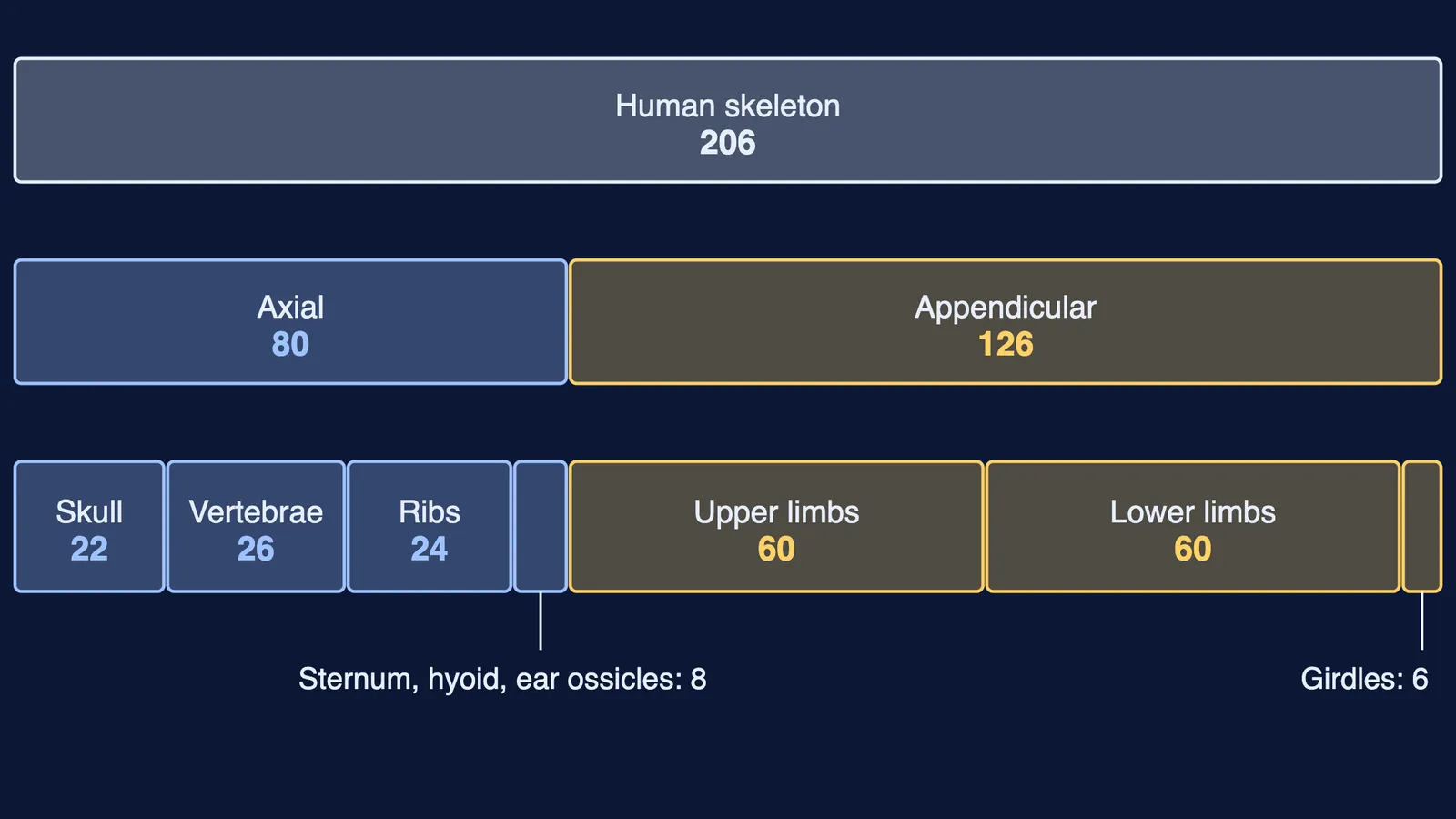
NCERT counts 206 bones in the human skeleton: 80 in the axial skeleton and 30 in each limb (NCERT Chapter 17).
Axial skeleton: bones along the body's main axis: skull, vertebral column, sternum and ribs. Don't confuse with: appendicular skeleton, the limbs and their girdles.
Atlas: the first of the 26 vertebrae; it meets the skull's two occipital condyles.
True, false and floating ribs: of the 12 pairs, pairs 1-7 reach the sternum through hyaline cartilage (true); 8-10 join the seventh rib (vertebrochondral or false); 11-12 have no front attachment (floating).
Pectoral girdle: clavicle and scapula on each side; the humerus fits into the glenoid cavity. Don't confuse with: pelvic girdle, two coxal bones, each a fusion of ilium, ischium and pubis, where the femur fits into the acetabulum.
What types of joints and disorders does the chapter cover?
Fibrous joint: an immovable joint of dense fibrous tissue. Example: sutures between skull bones.
Cartilaginous joint: bones linked by cartilage, allowing a little movement. Example: between adjacent vertebrae.
Synovial joint: a joint with a fluid-filled cavity, allowing wide movement. Examples: ball and socket (shoulder), hinge (knee), pivot (atlas and axis), gliding (between carpals), saddle (carpal and metacarpal of the thumb).
Myasthenia gravis: the body's immune system attacks the neuromuscular junction, so skeletal muscles tire, weaken and can become paralysed. Don't confuse with: muscular dystrophy, mostly genetic, gradual wasting of skeletal muscle.
Tetany: sudden, repeated muscle spasms caused by too little calcium (Ca++) in the body fluids.
Arthritis: inflamed joints, from any cause. Don't confuse with: gout, joint inflammation caused specifically by uric acid crystals.
Osteoporosis: bones lose mass with age and break more easily; falling estrogen levels are a common cause.
How to learn these terms
Draw one sarcomere and label Z, I, A, H and M. Redraw it contracted: I band and H zone shrink; A band stays.
Learn the pairs as pairs: troponin and tropomyosin, sarcolemma and sarcomere, Z line and M line, motor unit and neuromuscular junction.
Say the contraction chain from the callout aloud until you can start from any step.
Redraw the bone-count chart in the picture above from memory: 206 splits into 80 and 126, then into skull, vertebrae, ribs, limbs and girdles.
Nerve signals are covered in our Neural Control and Coordination Class 11 guide. If one term still will not stick, the Fermi Concept Library gives every concept in biology its own lesson and its place in the map.