

On this page
When you inspect muscle tissue on a histological slide or an exam diagram, three structural hallmarks tell the three muscle types apart: cell shape, striation banding, and the number and position of nuclei.
How do skeletal, smooth and cardiac muscle tissues differ under a microscope?
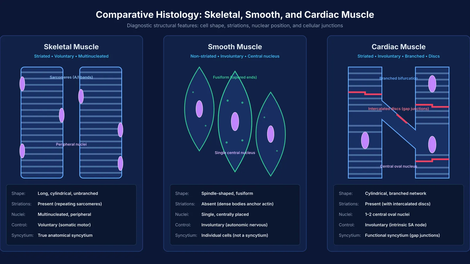
Skeletal muscle fibers are long, cylindrical, and strictly unbranched, ranging from 10 to 100 micrometers in diameter (McCuller et al., 2023). Under light microscopy, skeletal muscle displays alternate dark (anisotropic, or A-bands) and light (isotropic, or I-bands) striations produced by repeating sarcomere units (Campbell and Maani, 2023). Each fiber contains multiple oval nuclei situated at the periphery, pushed directly beneath the sarcolemma because the cell interior is packed with myofibrils.
Smooth muscle tissue, by contrast, is composed of unbranched, spindle-shaped cells that taper at both ends, a morphology termed fusiform (Hafen and Burns, 2023). Smooth muscle shows no cross-striations because actin and myosin filaments are not organized into sarcomeres; instead, thin actin filaments anchor into protein structures called dense bodies scattered throughout the cytoplasm and plasma membrane. Each smooth muscle cell contains only one oval nucleus located squarely in the center of its widest section.
Cardiac muscle fibers present a hybrid architecture. Like skeletal muscle, cardiac myocytes are cylindrical and striated, but they are shorter and branched, forming an interconnected, three-dimensional network (Arackal and Alsayouri, 2023). Each cardiac cell typically contains a single centrally placed nucleus, though occasionally two nuclei occur. The defining histological feature of cardiac muscle is the presence of intercalated discs—dense, stepped transverse bands that represent specialized cell junctions where plasma membranes interlock.
Why is cardiac muscle involuntary despite having striations?
Students frequently assume that having striations implies conscious voluntary control. In animal physiology, striations reflect mechanical organisation, not nervous regulation.
Striations simply show that actin and myosin are arranged in repeating sarcomeric units to generate rapid linear tension. Control over that machinery depends entirely on how the tissue receives its electrical signals. Skeletal muscle relies on somatic motor neurons terminating at discrete neuromuscular junctions; an action potential releases acetylcholine to initiate contraction, meaning you consciously recruit these fibers to run, write, or lift. In healthy adults, skeletal muscle accounts for approximately 40% of total body weight and contains 50% to 75% of all body protein (McCuller et al., 2023).
Cardiac muscle, although striated, is governed by intrinsic automaticity. Specialized autorhythmic cells in the sinoatrial node spontaneously depolarize, generating electrical impulses that spread across the myocardium without input from the conscious brain (Arackal and Alsayouri, 2023). While the autonomic nervous system modulates heart rate and contractile force through sympathetic and parasympathetic fibers, it does not initiate contractions.
How can you identify unknown muscle tissue in NEET questions?
In NEET-UG and Class 11 board examinations, questions on structural organisation in animal tissues often ask you to identify a tissue from an unlabeled diagram, or to match histological characteristics across multi-statement items. Mastering reading NCERT lines for NEET biology requires a reliable diagnostic order.
You can classify any muscle sample in four quick steps:
Check for striations: If striations are absent, the tissue is smooth muscle. If alternating dark and light bands are present, the tissue is either skeletal or cardiac muscle.
Inspect fiber branching: If the fibers branch and re-join, the tissue is cardiac muscle. If the fibers remain straight, cylindrical, and parallel without branching, it is skeletal muscle.
Locate the nuclei: If nuclei are multiple and pressed against the outer edge (periphery), the tissue is skeletal muscle. If the nucleus is single and centrally placed, it is smooth muscle (if non-striated) or cardiac muscle (if striated).
Search for intercalated discs: Dark, transverse zigzag lines marking cellular junctions definitively confirm cardiac muscle tissue.
When tackling assertion-reason questions where structural details blur together, building systematic conceptual clarity is essential. The Fermi Concept Library covers every concept in physics, chemistry, biology, mathematics and computing, each with its own lesson and its place in the map. If you encounter complex practice problems where multi-step statements seem contradictory, the Fermi Problem Library provides worked problems with a step-by-step solver that explains the reasoning rather than printing an answer.
What are the most common exam traps with muscle histology?
Examiners repeatedly target four classic points of confusion. Being aware of them helps you avoid losing marks when eliminating options in NEET biology MCQs.
The "striated equals voluntary" fallacy: NEET questions often state "all striated muscles are voluntary" as an option. This statement is false because cardiac muscle is striated yet strictly involuntary.
The esophageal muscle gradient: The muscularis externa of the human esophagus transitions along its length (Yasir and Menezes, 2023). The upper third contains striated skeletal muscle, the middle third contains a blend of skeletal and smooth muscle, and the lower third consists entirely of smooth muscle. Despite the skeletal histology of the upper third, swallowing becomes an involuntary reflex once food passes the pharynx.
The syncytium trap: Smooth muscle and cardiac muscle are never anatomical syncytia. Calling cardiac muscle a "syncytium" is only correct when specifying a functional syncytium, made possible by gap junctions that transmit ionic currents between adjacent cardiomyocytes (Arackal and Alsayouri, 2023).
Fatigue and energy expenditure: Skeletal muscle fatigues comparatively quickly when anaerobic glycolysis produces metabolic by-products. In contrast, cardiac muscle never fatigues under physiological conditions due to high mitochondrial density and rich vascularity (Arackal and Alsayouri, 2023), and smooth muscle maintains prolonged tone with minimal energy expenditure via its specialized latch state (Hafen and Burns, 2023).


