
On this page
Here are three original case studies on Anatomy of Flowering Plants, Chapter 6 of NCERT Class 11 Biology (Reprint 2026-27), each checked against that chapter. The CBSE 2026-27 Biology curriculum lists the chapter as "anatomy and functions of tissue systems in dicots and monocots". Treat these as practice in the case-based style.
New to the format? Read our guide to answering case-based questions. For the chapter, start with the Anatomy of Flowering Plants Class 11 guide.
Case 1: Counting stomata in Lucknow
In a Lucknow school lab, Ananya peels the epidermis from both surfaces of two leaves: Leaf X, a garden shrub's leaf with a net of veins, and Leaf Y, a grass blade with parallel veins. She counts stomata in five microscope fields per peel. Leaf X gives 18, 21, 17, 20, 19 (lower surface) and 3, 2, 4, 1, 5 (upper). Leaf Y gives 11, 9, 10, 12, 8 (upper) and 12, 10, 11, 13, 9 (lower). Leaf Y's guard cells are narrow in the middle and swollen at both ends.
(i) What shape are Leaf Y's guard cells? (a) Bean-shaped (b) Dumb-bell shaped (c) Barrel-shaped (d) Spherical
(ii) Find the mean number of stomata per field on each surface of Leaf X. How many times more stomata does the lower surface carry?
(iii) What makes up the stomatal apparatus? Which guard cell walls are highly thickened?
(iv) Which leaf is dorsiventral and which is isobilateral? Give two clues from the passage and one internal difference.
Answers.
(i) (b) Dumb-bell shaped, as in grasses.
(ii) Lower: (18 + 21 + 17 + 20 + 19) / 5 = 95 / 5 = 19 per field. Upper: (3 + 2 + 4 + 1 + 5) / 5 = 15 / 5 = 3 per field. The lower surface carries 19 / 3 ≈ 6.3 times as many.
(iii) The stomatal aperture, guard cells and surrounding subsidiary cells. The inner walls, towards the pore, are highly thickened.
(iv) Leaf X is dorsiventral: reticulate venation and far more stomata on the abaxial (lower) surface. Leaf Y is isobilateral: parallel venation and stomata on both surfaces (means 10 and 11). Inside, X's mesophyll has palisade and spongy parenchyma; Y's is undifferentiated.
Case 2: Which root slide is the monocot?
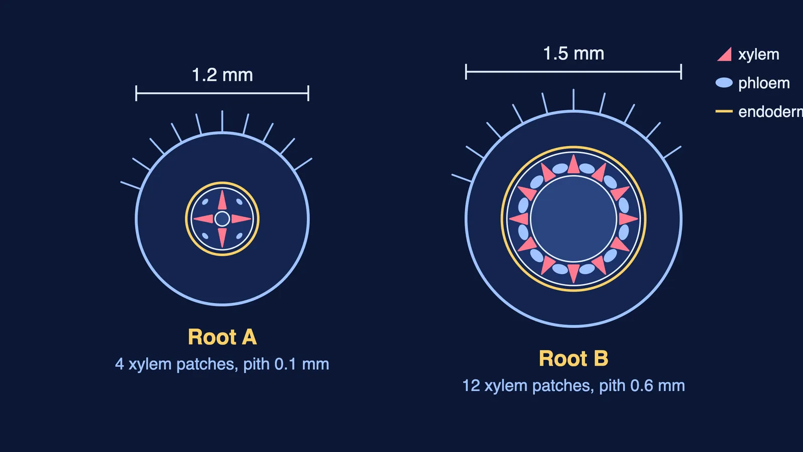
In a Kochi practical, Rahul gets two slides of young root transverse sections, marked A and B. Root A is 1.2 mm across, with 4 xylem patches alternating with 4 phloem patches and a pith only 0.1 mm wide. Root B is 1.5 mm across, with 12 xylem patches alternating with 12 phloem patches and a pith 0.6 mm wide. In both, a single ring of barrel-shaped cells with no intercellular spaces, and dark bands on their walls, surrounds the centre. In Root A, a small bulge is pushing outward from a layer just inside that ring.
(i) How are xylem and phloem arranged in both roots? (a) Conjoint open (b) Conjoint closed (c) Radial (d) Scattered
(ii) Express each pith width as a percentage of the root's diameter. Which root is a monocot?
(iii) Name the ring of barrel-shaped cells, the dark bands and the substance deposited in them.
(iv) From which layer is Root A's bulge arising, and what is it? Which tissues together make up the stele?
Answers.
(i) (c) Radial: xylem and phloem alternate on different radii, as in roots.
(ii) Root A: 0.1 / 1.2 × 100 ≈ 8.3%. Root B: 0.6 / 1.5 × 100 = 40%. Root B is the monocot: it is polyarch (more than six xylem bundles) with a large, well-developed pith. Root A is a dicot.
(iii) The endodermis. The bands are casparian strips of suberin, a waxy, water-impermeable material.
(iv) The pericycle, a few layers of thick-walled parenchymatous cells just inside the endodermis; a lateral root is being initiated. The stele is everything inside the endodermis: pericycle, vascular bundles and pith.
Case 3: A dry spell on a farm near Jaipur
Near Jaipur, a farmer grows maize beside sunflowers. In a dry spell, an agriculture student tags 40 maize leaves. At 7 am none are curled; at 1 pm, 34 have rolled inwards. The field is irrigated at 3 pm, and by 5 pm only 6 are still curled. She also cuts sections of young stems from both crops. Section P shows vascular bundles in a ring, with semi-lunar patches of sclerenchyma above the phloem. Section Q shows many scattered bundles, each wrapped in a sclerenchyma sheath, with water-containing cavities inside.
(i) Which cells make the maize leaves curl? (a) Guard cells (b) Bulliform cells (c) Subsidiary cells (d) Bundle sheath cells
(ii) Find the percentage of tagged leaves curled at 7 am, 1 pm and 5 pm. Explain the change after irrigation in one line.
(iii) Which section is from the sunflower? Name the type of vascular bundle it has.
(iv) Are Section Q's bundles open or closed? Say what decides this, and give two other features that mark Q as a monocot stem.
Answers.
(i) (b) Bulliform cells: large, empty, colourless adaxial epidermal cells along the veins.
(ii) 7 am: 0 / 40 = 0%. 1 pm: 34 / 40 × 100 = 85%. 5 pm: 6 / 40 × 100 = 15%. Irrigation lets the bulliform cells absorb water and turn turgid, so the leaves unroll; when flaccid under water stress, they curl leaves inwards to cut water loss.
(iii) Section P: a ring of bundles and semi-lunar sclerenchyma patches of pericycle mark a dicot stem. Each bundle is conjoint, open, with endarch protoxylem.
(iv) Closed. Cambium decides it: P's bundles have cambium between phloem and xylem, while Q's (maize, a monocot) have none. Q's other monocot features: scattered bundles, each wrapped in a sclerenchymatous bundle sheath, and water-containing cavities in them.
How do you attack case-study questions on plant anatomy?
Sort clues first: dicot or monocot, then root, stem or leaf.
Endodermis is the innermost cortex layer; pericycle lies just inside it.
Link "open" and "closed" to cambium, never to the bundle sheath.
In data parts, show each step, then say what the number means.
Learn the grass features as a pair: dumb-bell guard cells and bulliform cells.
For more practice, try our assertion-reason questions on anatomy of flowering plants. If the layers still blur together, the Fermi Concept Library covers every concept in biology, each with its own lesson.