
On this page
What do case-study questions on Cell: The Unit of Life test?
Cell: The Unit of Life is Chapter 8 of NCERT Class 11 Biology (Reprint 2026-27). The CBSE Class XI Biology curriculum for 2026-27 places it in Unit III, Cell: Structure and Function, which carries 15 of 70 theory marks along with Biomolecules and Cell Cycle and Cell Division. Class 11 papers are set by your school, so treat these as practice in the case-based style, not a board pattern.
The three cases below are ours, easy to hard. Revise first with our Class 11 guide to Cell: The Unit of Life.
Case 1: Onion peel and cheek cells in a Lucknow lab
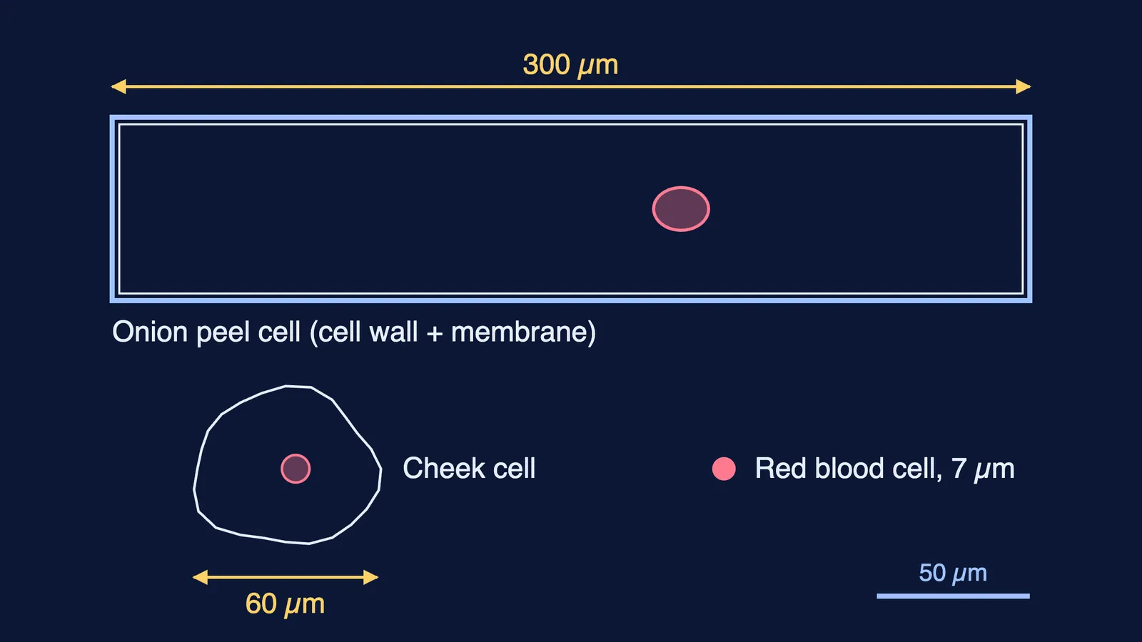
In a Class 11 practical in Lucknow, Meera mounts a stained onion peel and a smear of her own cheek cells. Her eyepiece micrometer has been calibrated so that, at this magnification, one division equals 2.5 µm. A typical onion cell spans 120 divisions along its length; a cheek cell spans 24 divisions across. Both cells show a dense, stained nucleus. The onion cells have a thick outer line; the cheek cells are flat and irregular. Neither slide shows mitochondria or ribosomes, though both cells have them.
(i) What forms the outer boundary of the cheek cell? (a) Cell wall (b) Plasma membrane (c) Middle lamella (d) Tonoplast
(ii) Both cells have a membrane-bound nucleus. What type of cell does that make them?
(iii) Find the length of the onion cell and the width of the cheek cell in µm. How many times longer is the onion cell?
(iv) Why can Meera see neither mitochondria nor ribosomes on her slides?
Answers.
(i) (b). NCERT says cheek cells are bounded by an outer membrane; only the onion cell has a wall.
(ii) Eukaryotic.
(iii) Onion: 120 × 2.5 = 300 µm. Cheek: 24 × 2.5 = 60 µm. 300 ÷ 60 = 5 times.
(iv) NCERT notes that mitochondria are not easily visible under the microscope unless specifically stained. Ribosomes were first observed under the electron microscope, by George Palade in 1953, so a light microscope will not show them.
Case 2: Two kinds of secretory cell in a Hyderabad lab
A research team in Hyderabad compares electron micrographs of two animal cells. Cell A secretes digestive enzymes, which are proteins. Cell B makes a steroid hormone. They lay a grid over each cell's cytoplasm and record the structure under each of 500 points. Cell A: rough ER 165, smooth ER 10, Golgi 30, mitochondria 40, rest 255. Cell B: rough ER 20, smooth ER 140, Golgi 15, mitochondria 60, rest 265. Cell A also has unusually large and numerous nucleoli.
(i) What makes rough ER "rough"? (a) Cisternae stacked in piles (b) Ribosomes attached to its outer surface (c) Pores in its membrane (d) Folds called cristae
(ii) What percentage of the cytoplasm is rough ER in Cell A, and smooth ER in Cell B?
(iii) Which cell makes the steroid hormone? Use the data and the function of each kind of ER.
(iv) Explain why Cell A has large, numerous nucleoli.
Answers.
(i) (b). NCERT calls ER bearing ribosomes rough ER.
(ii) Cell A: 165 ÷ 500 × 100 = 33%. Cell B: 140 ÷ 500 × 100 = 28%.
(iii) Cell B. Its smooth ER (28%) outweighs its rough ER (4%) by 7 times, and NCERT says smooth ER is the major site of lipid synthesis; in animal cells, lipid-like steroidal hormones are made there.
(iv) The nucleolus is a site of active ribosomal RNA synthesis, and NCERT notes that cells actively making proteins have larger and more numerous nucleoli. Cell A needs many ribosomes for its rough ER.
Case 3: Mitochondria in two cell lines in Patiala
A college lab in Patiala studies electron micrographs of two cultured animal cell lines, P and Q. In P, the students count 24 mitochondria per 100 µm² of cytoplasm; in Q, 8. In a longitudinal section of a typical mitochondrion, the outer membrane traces to 5.0 µm in both; the inner membrane, with its folds, traces to 15.0 µm in P and 7.5 µm in Q. A mitochondrion in P measures 0.6 µm wide and 2.2 µm long. Arjun argues that because mitochondria are membrane-bound, they must be part of the endomembrane system and are made by the ER.
(i) Where in a mitochondrion are its circular DNA and 70S ribosomes? (a) Outer membrane (b) Inter-membrane space (c) Matrix (d) Cristae surface
(ii) Is P's mitochondrion within the size range NCERT gives? Show the comparison.
(iii) Compare P and Q using both the counts and the membrane tracings. Which cell line is likely more active in aerobic respiration?
(iv) Evaluate Arjun's argument.
Answers.
(i) (c). NCERT places a single circular DNA molecule and 70S ribosomes in the matrix.
(ii) Yes. NCERT gives a diameter of 0.2-1.0 µm and a length of 1.0-4.1 µm; 0.6 µm and 2.2 µm both fall inside.
(iii) P has 24 ÷ 8 = 3 times as many mitochondria. Its inner-to-outer membrane ratio is 15.0 ÷ 5.0 = 3, against 7.5 ÷ 5.0 = 1.5 for Q, so it has more cristae. Cristae increase surface area, and mitochondrial number varies with physiological activity, so P is likely more active in aerobic respiration.
(iv) Wrong. NCERT keeps mitochondria, chloroplasts and peroxisomes out of the endomembrane system because their functions are not coordinated with the ER, Golgi, lysosomes and vacuoles. Mitochondria also divide by fission.
How do you answer case-study questions on this chapter?
Convert units first. Divisions to µm, nm to µm (1 µm = 1,000 nm).
Turn counts into percentages or ratios, one line of working per step.
Link structure to function in one sentence. "More cristae, more surface area for aerobic respiration" earns more than "more cristae".
Know the outsiders. Mitochondria, chloroplasts and peroxisomes are not endomembrane organelles; ribosomes have no membrane.
Know what a light microscope shows. Mitochondria need staining; ribosomes need an electron microscope.
For the general method, read our guide to case-based questions in board exams, then test your reasoning with the Cell: The Unit of Life assertion-reason questions. If an organelle feels shaky, the Fermi Concept Library has every concept in physics, chemistry, biology, mathematics and computing, each with its own lesson.