
On this page
What do body fluids and circulation case-study questions test?
Each case below is a short passage followed by four questions of rising difficulty. Class 11 exams are set by your school, so treat these as practice in the case-based style.
The CBSE Biology curriculum for 2026-27 lists Chapter 15, Body Fluids and Circulation, from blood groups and clotting to the cardiac cycle, ECG and circulatory disorders. The cases and numbers are ours; every fact follows the NCERT Class 11 Biology chapter (Reprint 2026-27).
For the chapter in brief, read our Body Fluids and Circulation Class 11 guide. For the general method, see how to handle case-based questions.
Case 1: Which blood can a patient receive?
A college NSS unit in Pune collects 120 units at a one-day blood donation camp. The tally by group reads: O+ 40, O− 6, B+ 34, B− 4, A+ 23, A− 3, AB+ 9 and AB− 1. That evening a hospital asks for blood for an accident patient of group A, Rh negative. Meera, a volunteer, is Rh negative and her husband Rh positive; her doctor has planned an injection for her after their first child is born.
(i) The plasma of a group B donor contains: (a) anti-A (b) anti-B (c) both anti-A and anti-B (d) neither
(ii) Which ABO group in the tally can give blood to a person of any ABO group, and what are such people called?
(iii) How many of the camp's 120 units can safely go to the hospital's patient?
(iv) Why does Meera's doctor plan an injection after her first delivery, and what would it contain?
Answers.
(i) (a). Group B has antigen B on its RBCs and anti-A antibodies in its plasma.
(ii) Group O, whose RBCs carry neither A nor B antigen. They are called universal donors.
(iii) An A patient can take A or O blood, and an Rh-negative person exposed to Rh-positive blood forms antibodies against the Rh antigen, so only Rh-negative units work: A− 3 + O− 6 = 9 units.
(iv) If the baby is Rh positive, foetal blood may reach hers during delivery and she starts making anti-Rh antibodies, which in a later pregnancy can destroy an Rh-positive foetus's RBCs (erythroblastosis foetalis). Anti-Rh antibodies given right after the first delivery avoid this.
Case 2: How hard does an athlete's heart work?
Riya, 17, trains for the 400 m at a sports academy in Bengaluru. At rest her heart rate is 60 beats per minute and an echo test puts her stroke volume at 80 mL. In the final straight of a time trial, her heart rate reaches 150 beats per minute and her stroke volume 110 mL. Her coach points out that the heart beats on its own, yet it speeds up when they race.
(i) The part of the heart that sets Riya's resting rate is the: (a) AV node (b) bundle of His (c) SA node (d) Purkinje fibres
(ii) How long does one cardiac cycle last at rest and during the run?
(iii) Calculate Riya's cardiac output at rest and during the run. By what factor does it rise?
(iv) The heart is myogenic. Explain how Riya's heart rate and output still rise during the race.
Answers.
(i) (c). The SA node generates the most action potentials per minute: it is the pacemaker.
(ii) At rest: 60 s ÷ 60 beats = 1.0 s per cycle. During the run: 60 s ÷ 150 beats = 0.4 s per cycle.
(iii) Cardiac output = stroke volume × heart rate. Rest: 80 mL × 60 = 4,800 mL, or 4.8 L per minute. Run: 110 mL × 150 = 16,500 mL, or 16.5 L per minute. The rise is 16.5 ÷ 4.8 ≈ 3.4 times.
(iv) Nodal tissue starts each beat, but a centre in the medulla oblongata moderates it through the autonomic nervous system. Sympathetic signals raise the heart rate and the strength of ventricular contraction; adrenal medullary hormones also increase cardiac output.
Case 3: What does a grandfather's ECG strip show?
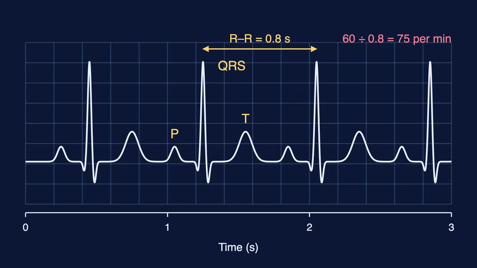
Mr Verma, 68, visits a Lucknow clinic with chest pain on climbing stairs that eases at rest. A technician records a standard ECG with leads on both wrists and the left ankle. The paper moves at 25 mm per second, and the distance between two R peaks is 20 mm. His blood pressure on three visits reads 150/95, 148/94 and 152/96 mm Hg. Tests show deposits narrowing the arteries to his heart muscle. The cardiologist says it is not heart failure.
(i) The QRS complex in his ECG represents: (a) atrial depolarisation (b) ventricular depolarisation (c) ventricular repolarisation (d) closure of the semilunar valves
(ii) Calculate his heart rate from the strip.
(iii) Do his readings show hypertension? Name the two numbers in 150/95.
(iv) Name his condition and his symptom, then explain how heart failure differs from both a heart attack and cardiac arrest.
Answers.
(i) (b). P is atrial depolarisation; T is ventricular repolarisation.
(ii) R-R interval = 20 mm ÷ 25 mm per s = 0.8 s. Heart rate = 60 ÷ 0.8 = 75 beats per minute.
(iii) Yes. Repeated readings of 140/90 or higher show hypertension. In 150/95, 150 mm Hg is the systolic (pumping) pressure and 95 mm Hg is the diastolic (resting) pressure.
(iv) Coronary arteries narrowed by deposits of calcium, fat, cholesterol and fibrous tissue mean coronary artery disease (atherosclerosis); chest pain when too little oxygen reaches the heart muscle is angina. Heart failure is a heart not pumping effectively enough for the body's needs; a heart attack is sudden damage to heart muscle from poor blood supply; cardiac arrest is the heart stopping.
How to attack case-study questions
List what the passage gives (rates, volumes, groups, readings) before you answer.
For blood-group cases, check ABO first and Rh second, and count only units that pass both.
In heart numericals, write the formula first (cardiac output = stroke volume × heart rate) and keep units on every line.
Match each ECG wave to one event: P with atria, QRS with ventricles contracting, T with ventricles returning to rest.
For disorders, use NCERT's exact terms and say what each one is not.
If one idea keeps tripping you up, the Fermi Concept Library has a lesson for every concept in biology.