
On this page
Lots of students can name the four chambers of the heart, then freeze when a question asks where blood goes after the pulmonary veins. The fix is to stop memorising a list and follow one traveller. Below, we trace one red blood cell through both circuits.
What is double circulation in humans?
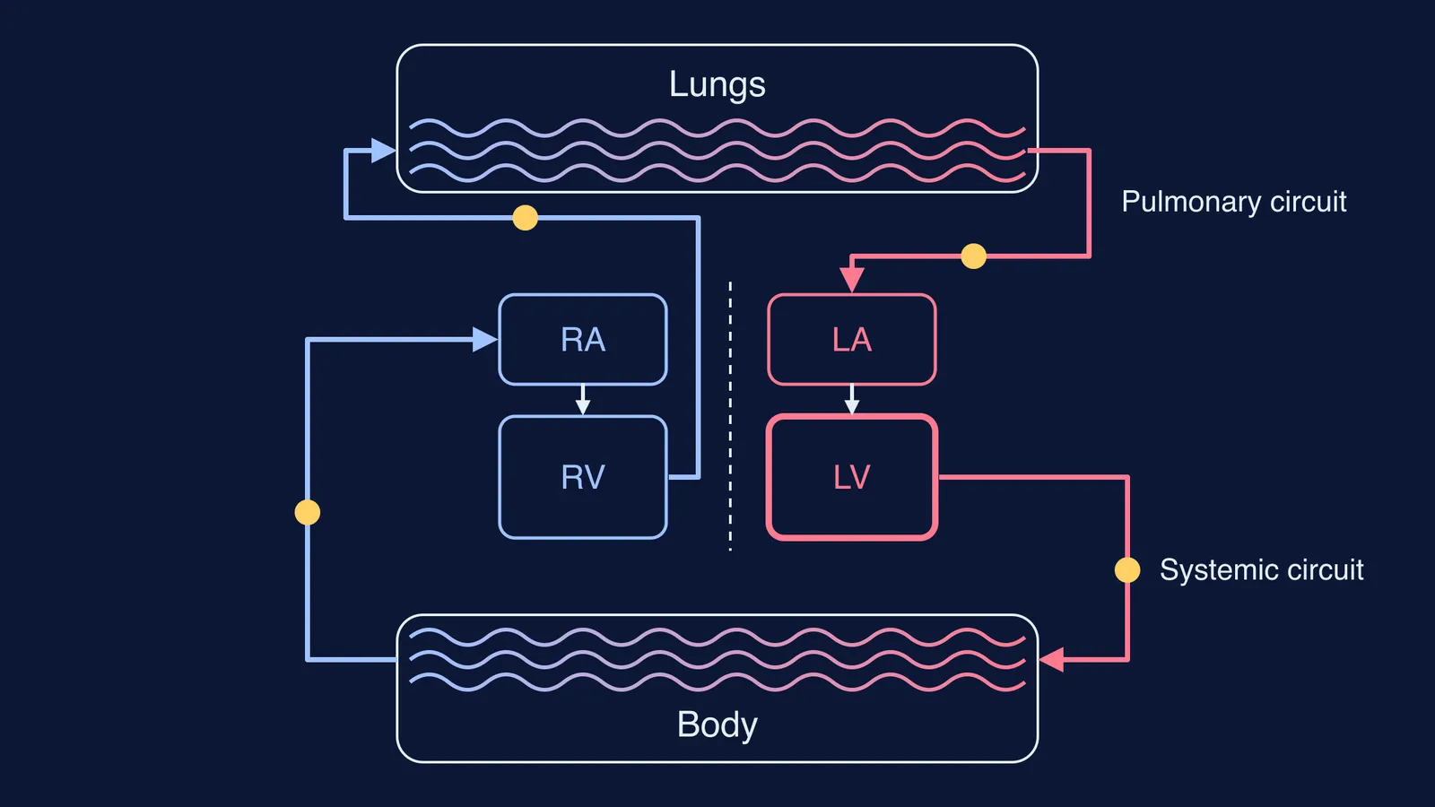
Your blood runs through two circuits joined at the heart: the pulmonary circuit, where the right ventricle pumps blood to the lungs for gas exchange, and the systemic circuit, which carries oxygen-rich blood to the rest of the body (OpenStax Anatomy and Physiology 2e, section 20.5). The two circuits are joined one after the other, so any single cell passes through the heart twice on every full lap.
Meet our traveller. A red blood cell has a mean diameter of only about 7 to 8 micrometres, according to OpenStax Anatomy and Physiology 2e, section 18.3. It may hold about 300 million haemoglobin molecules, and each haemoglobin molecule can carry four oxygen molecules. It lives up to 120 days.
What is the path of blood through the heart, step by step?
We start our cell in a capillary in your big toe, where it has just handed over its oxygen. The route below follows OpenStax Anatomy and Physiology 2e, section 19.1.
Toe capillaries to small veins, then the inferior vena cava (blood from the head and arms uses the superior vena cava).
Into the right atrium.
Through the tricuspid valve into the right ventricle.
Through the pulmonary valve into the pulmonary trunk, which splits into the left and right pulmonary arteries.
Into the lung capillaries: carbon dioxide leaves, oxygen loads onto haemoglobin.
Back through the pulmonary veins (four of them, two on the left and two on the right, per OpenStax section 20.5) into the left atrium.
Through the mitral (bicuspid) valve into the left ventricle.
Through the aortic valve into the aorta, then out through arteries to capillaries all over the body.
Steps 2 to 4 are the first pass through the heart (right side, oxygen-poor). Steps 6 to 8 are the second pass (left side, oxygen-rich).
Why do mammals keep oxygenated and deoxygenated blood separate?
It helps to compare hearts across animals, as OpenStax Biology 2e, section 40.1 does:
Fish have a two-chambered heart. Blood goes heart, gills, body, in one loop. The textbook says this limits the oxygen that reaches some organs and reduces the overall metabolic capacity of fish.
Amphibians have three chambers: two atria and a single ventricle. Some blood mixes in that ventricle, which reduces the efficiency of oxygenation.
Mammals and birds have four chambers that completely separate oxygenated and deoxygenated blood. The same source says this is probably required for the warm-blooded lifestyle of mammals and birds.
Separation brings two gains. First, the blood the left ventricle sends to the body is not diluted with oxygen-poor blood, which is why the textbook ties it to warm-bloodedness. Second, the two sides can work at different pressures. In OpenStax section 19.1, the left ventricle has much thicker muscle because it must push blood round the whole body, while the right ventricle needs less pressure because the lung circuit is shorter and offers less resistance. Even so, both ventricles pump the same amount per beat, normally 70 to 80 mL (OpenStax section 19.3).
Compare all the classes in our guide to heart chambers across vertebrate classes.
How do you trace blood flow in an exam answer?
When a question drops you somewhere in the loop, ask three questions in order:
Which side of the heart is this on? In an adult, right means oxygen-poor and left means oxygen-rich.
Is the vessel heading towards the heart (vein) or away from it (artery)?
Which valve did the blood just cross, and which one comes next?
Worked example. A cell is in the aorta. How many heart valves does it cross before it next reaches the lungs? Walk it: aorta, body capillaries, vena cava, right atrium, tricuspid valve, right ventricle, pulmonary valve, pulmonary artery, lungs. Answer: two valves, the tricuspid and the pulmonary (the aortic valve is already behind it).
To practise, sketch the loop as a figure of eight from memory, lungs on top and body below, then talk one cell round it out loud. If valve names trip you up, our guide to heart valve names and positions pairs each valve with the chambers on either side.
Where can you see double circulation interactively?
A flat diagram hides the fact that this loop never stops. The Fermi Simulation Lab has interactive simulations for the ideas that are easier to see than to read, and the Fermi Concept Library gives every concept in biology its own lesson and its place in the map, so you can connect circulation to respiration and blood.
For a shorter recap of the same topic, see double circulation in humans explained.