

On this page
The heart is a mechanical pump. To keep blood moving forward and never backward, it relies on a system of one-way doors. If you are studying human physiology, getting the positions of the tricuspid, bicuspid and semilunar valves straight is a guaranteed way to secure marks.
Here is how the valves are arranged, what the chordae tendineae do, and how to remember the left and right circuits without mixing them up.
Where are the atrioventricular (AV) valves?
According to anatomical reviews of the heart, a normal human heart uses exactly four valves. The atrioventricular valves sit between the upper chambers (atria) and the lower chambers (ventricles). Their job is to prevent blood from flowing back up into the atria when the powerful ventricles contract. (While the AV valves physically separate these chambers, the electrical signal that coordinates their beat travels through a separate structure discovered by Sunao Tawara and the atrioventricular node of the heart).
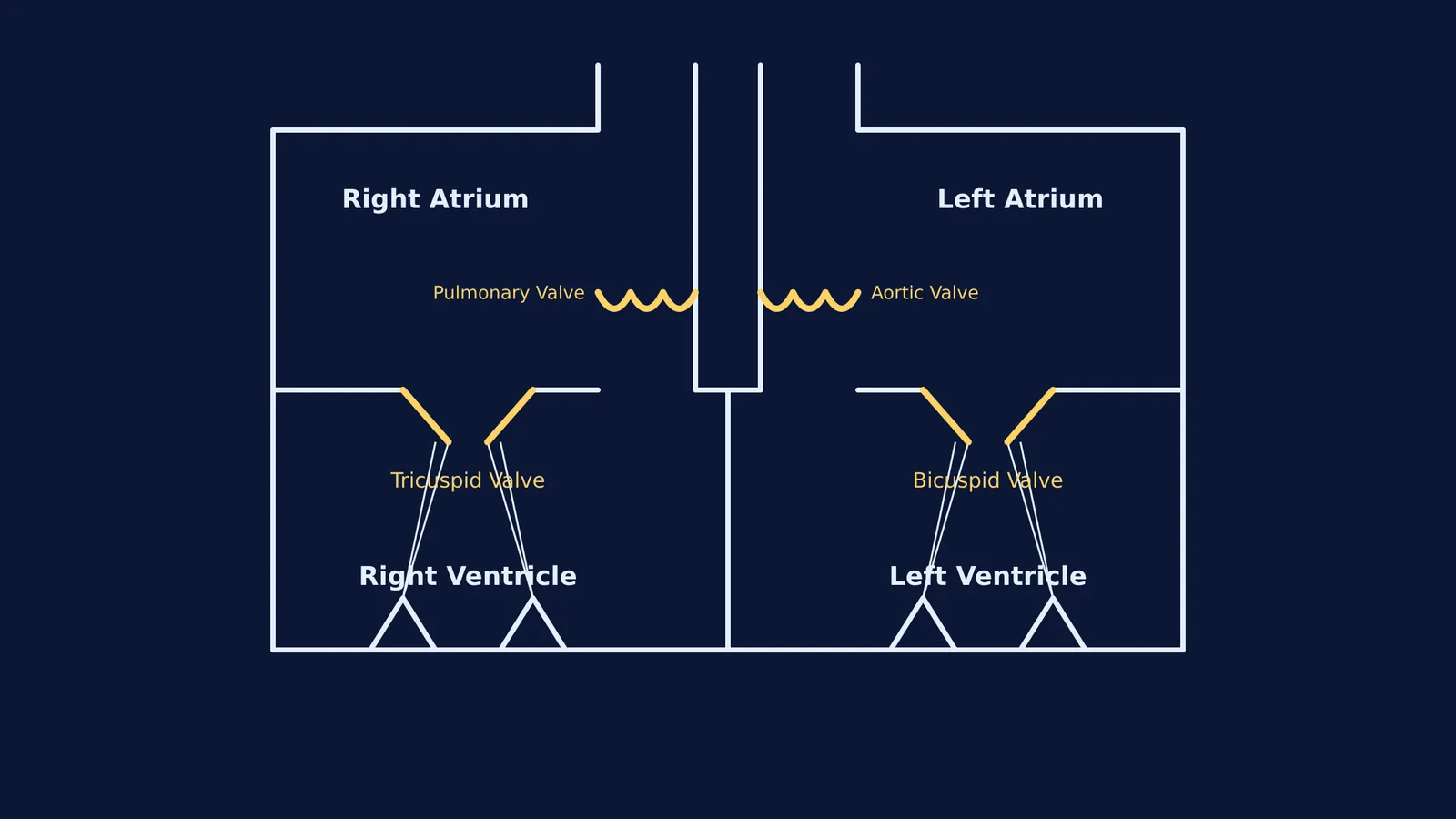
Tricuspid valve: Located between the right atrium and the right ventricle. As its name suggests, it consists of three muscular flaps (cusps).
Bicuspid (mitral) valve: Located between the left atrium and the left ventricle. It is built from exactly two flaps.
If you keep mixing up the left and right circuits, the Concept Library lays out the entire human circulatory system, each idea with its own lesson and its place in the map.
What do the chordae tendineae do?
The chordae tendineae, often called the "heart strings", are tough, fibrous cords of connective tissue. They attach the flaps of the tricuspid and bicuspid valves to the papillary muscles on the inner walls of the ventricles.
When the ventricles contract to push blood out of the heart, the pressure inside them spikes. The papillary muscles contract simultaneously, pulling the chordae tendineae taut. This tension anchors the valve flaps, preventing them from blowing backwards (everting) into the atria—much like the spokes of an umbrella stopping it from flipping inside out in a strong wind.
Visualising how the papillary muscles pull the strings at exactly the right moment is easier when you see it in motion. The Fermi Simulation Lab has interactive simulations for the ideas that are easier to see than to read, letting you explore a beating heart to observe the timing of each contraction.
Where are the semilunar valves?
While the AV valves guard the entrances to the ventricles, the semilunar valves guard the exits. They are located at the bases of the large arteries leaving the heart, and they prevent blood from falling back into the ventricles once it has been pumped out. Both semilunar valves are built with three half-moon-shaped cusps, as detailed in anatomical reviews.
Pulmonary semilunar valve: Positioned between the right ventricle and the pulmonary artery.
Aortic semilunar valve: Positioned between the left ventricle and the aorta.
Importantly, the semilunar valves do not have chordae tendineae. Their cup-like shape catches the falling blood when the ventricles relax, snapping them shut automatically.
How does blood flow through the heart?
A common exam question asks you to trace a drop of blood through the heart. You can map it out using the valves as checkpoints—a physiological map understood centuries before Christiaan Barnard and the first human heart transplant made headlines:
Deoxygenated blood enters the right atrium and passes through the tricuspid valve into the right ventricle.
The right ventricle pumps it through the pulmonary semilunar valve into the pulmonary artery, heading to the lungs—the circuit first described by Ibn al-Nafis and the flow of blood through the lungs.
Oxygenated blood returns to the left atrium and drops through the bicuspid valve into the left ventricle.
The left ventricle pumps it fiercely through the aortic semilunar valve into the aorta, sending it to the rest of the body.
If you are ever stuck on a question about why the bicuspid valve needs thicker strings or why the left ventricle wall is so thick, you can ask Fermi's AI tutor, which works through problems alongside the student, to explain the pressure differences step by step.


