
On this page
Cut a molar in half from top to bottom and you see layers. A tooth-structure question asks you to name those layers and say what each does. Here they are from the outside in, with a labelled section and a checklist for drawing it in an exam.
What are the main parts of a tooth?
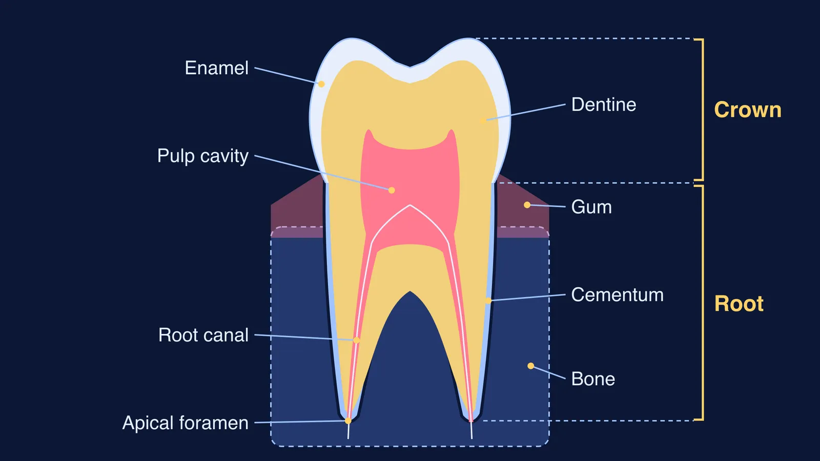
The crown is the visible part of the tooth, normally covered by enamel, according to the American Dental Association's MouthHealthy guide. The root sits below the gum, in a socket in the jaw bone. The Charles University histology textbook names three parts: crown, neck and root, with the neck where the other two meet.
Four tissues build them:
Enamel covers the crown.
Dentine lies under the enamel and the cementum.
Pulp fills the hollow centre.
Cementum covers the root.
The diagram above is a labelled section of a two-rooted molar. Enamel stops at the neck, and cementum starts there. That detail is the easiest one to get wrong.
Why is enamel so hard, and why can it not regrow?
Enamel is the hardest tissue in the human body, and in adults mineral makes up as much as 96% of it, according to the Charles University textbook. A review in Comptes Rendus Chimie by Dessombz and colleagues puts it at 97% mineral, mostly the calcium phosphate hydroxyapatite, with the other 3 to 4% water and protein, and calls enamel the most mineralised biological tissue.
The same review explains the catch: enamel cannot regrow, because the cells that make it, the ameloblasts, are lost. The ADA guide puts it plainly: enamel has no living cells, so it cannot repair damage from decay or wear. That is why protecting it matters, as our post on why toothpaste contains fluoride explains with the acid and pH chemistry.
What is dentine, and how is it different from enamel?
Dentine is about 70% inorganic material, 20% organic material and 10% water, according to the Charles University textbook. So it is less mineralised and softer than enamel.
Dentine contains microscopic tubules, small hollow canals, as the ADA guide describes. Unlike enamel, it keeps its makers: the odontoblasts, the dentine-forming cells, line the edge of the pulp, according to an oral histology lecture from the University of Baghdad's College of Dentistry.
What is inside the pulp cavity?
The pulp is the soft, non-calcified centre of the tooth, containing nerves, blood vessels and connective tissue, says the ADA guide. The Baghdad histology lecture divides the space in two:
The pulp chamber in the crown holds the coronal pulp.
The root canal in each root holds the radicular pulp.
The root pulp joins the tissues outside the tooth through an opening at the root tip, the apical foramen. The same lecture notes that the pulp shrinks with age, because dentine keeps being laid down on its walls.
What holds the root in the jaw?
Cementum is the hard connective tissue covering the root, and it gives attachment to the periodontal ligament, according to the ADA guide. It is about 65% inorganic material, 23% organic material and 12% water, per the Charles University textbook, a little less mineralised than dentine.
How do you draw and label a tooth section for an exam?
Work in this order, so that no layer ends up in the wrong place.
Draw the outline: a crown with two cusps, two roots, and a mark at the neck.
Shade a thin enamel layer over the crown only, thickest at the cusps and thinning to nothing at the neck.
Draw the pulp cavity: a chamber in the crown with small horns toward the cusps, narrowing into one canal per root.
Run each canal to the root tip and label the opening the apical foramen.
Add a thin cementum line along the outside of the roots only.
Draw the gum around the neck and bone around the roots.
Label with straight lines that do not cross: crown, root, enamel, dentine, pulp cavity, root canal, cementum, gum, bone.
Then check for three mistakes:
Enamel drawn down over the root.
A pulp cavity that stops short of the root tip.
Dentine left unlabelled, though it runs through crown and root.
For the wider digestion chapter, see our labelled figure of the alimentary canal and our guide to the types of teeth and the dental formula. If you keep mixing up which tissue goes where, the Fermi Concept Library gives every biology concept its own lesson and its place in the map, so you can see how teeth connect to the rest of digestion.
A tooth that hurts, aches with hot or cold, or looks damaged needs a dentist, not a diagram.