On this page
When a question paper says "draw a neat, labelled diagram of the human alimentary canal", most marks go to two things: the parts in the right order, and the glands joined to the right place. The NCERT Class 10 Science chapter Life Processes describes the alimentary canal as "basically a long tube extending from the mouth to the anus" and shows it in Figure 5.6 (NCERT, Class 10 Science, Chapter 5). This guide gives you the full order, the glands, the labels students skip, and a way to draw it from memory.
What are the parts of the alimentary canal, in order?
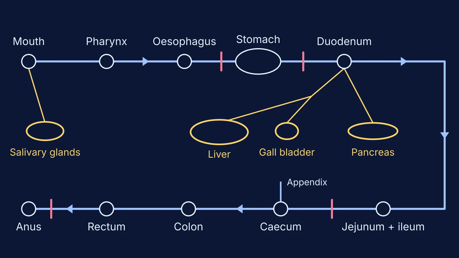
Follow the food. Each part below is a section of the same tube, and the order is taken from OpenStax Anatomy and Physiology, section 23.1 and the sections that follow it.
Mouth (buccal cavity), with teeth and tongue
Pharynx, the short throat tube shared with the airway
Oesophagus (food pipe)
Stomach: cardia, fundus, body and pylorus
Small intestine: duodenum, jejunum, ileum
Large intestine: caecum (with the appendix), colon, rectum, anal canal
Anus
The alimentary canal is about 7.6 metres (25 feet) long in a living adult and closer to 10.7 metres (35 feet) when measured after death (OpenStax 23.1). The small intestine is about 3 metres (10 feet) long in a living person, and its first part, the duodenum, is only about 25 cm (10 inches) (OpenStax 23.5).
Which digestive glands go on the labelled figure?
Some glands sit outside the tube and send juices in through ducts; others lie in its wall.
Salivary glands (three pairs): the parotid glands near the ears, the submandibular glands in the floor of the mouth and the sublingual glands below the tongue (OpenStax 23.3). Saliva contains salivary amylase, which breaks down starch (NCERT).
Gastric glands in the stomach wall release hydrochloric acid, the enzyme pepsin and mucus (NCERT).
Liver: the largest gland in the body, weighing about three pounds in an adult (OpenStax 23.6). It makes bile.
Gall bladder: stores and concentrates bile, then pushes it into the duodenum through the common bile duct (OpenStax 23.6).
Pancreas: secretes pancreatic juice with trypsin and lipase (NCERT), and also makes the hormones insulin and glucagon (OpenStax 23.6).
Intestinal glands in the wall of the small intestine secrete intestinal juice (NCERT).
Which parts do students forget to label?
Pharynx. Students jump straight from mouth to oesophagus. The pharynx runs from the back of the mouth and nose to the openings of the oesophagus and larynx (OpenStax 23.3).
Duodenum. It is the first part of the small intestine, and the main pancreatic duct joins the common bile duct just before both empty into it (OpenStax 23.6). Draw the ducts ending there, not in the stomach.
Sphincters. NCERT names the sphincter that lets food out of the stomach in small amounts and the anal sphincter (NCERT). OpenStax adds the lower oesophageal sphincter, the pyloric sphincter and the ileocaecal valve where the small intestine meets the large (OpenStax 23.4, 23.5).
Caecum and appendix. The appendix hangs off the caecum, the pouch at the start of the large intestine (OpenStax 23.5).
Rectum. NCERT's Figure 5.6 goes from colon to anus, but the rectum is the final 20 cm (8 inches) or so of the canal (OpenStax 23.5).
Diaphragm. NCERT shows it as a landmark that the oesophagus passes through. It is a muscle, not part of the canal, so do not list it as a digestive organ.
How do you draw the alimentary canal quickly?
Use this five-step order. Practise it three times this week, with the book closed, then compare with Figure 5.6.
Draw the head outline and a mouth, then a straight oesophagus down the centre of the chest.
Draw the stomach as a J-shape on the left side of the body (your right on the page), just below the diaphragm line.
Add the liver as a large wedge on the other side, with the small gall bladder tucked under it, and the pancreas as a leaf shape below the stomach.
Draw the duodenum from the stomach outlet, then the coiled small intestine in the centre, framed by the large intestine: up, across and down.
Finish with the appendix, rectum and anus, then label in the order food travels, glands last.
If the drawing is fine but the reason behind it is shaky, such as why bile must make the food alkaline before pancreatic enzymes work, read the digestion lessons in the Fermi Concept Library, then try questions on them in the Fermi Problem Library, which explains the reasoning step by step.
For more practice, see how to revise using textbook diagrams with the cover-and-check method and why your stomach does not digest itself: the mucus barrier. To see what the digestive enzymes are doing chemically, read how enzymes lower activation energy on a graph.