

On this page
Why does gastric acid not burn through the stomach wall?
Every day, the human stomach secretes roughly two litres of gastric juice containing concentrated hydrochloric acid at concentrations up to 150 mM, maintaining an average luminal pH between 1.5 and 2.0, as detailed in Adrian Allen and Gunnar Flemström's physiological review. This acidic environment dissolves dense food particles and destroys ingested pathogens. Alongside acid, the stomach secretes pepsin, a powerful endopeptidase that digests proteins into peptide fragments.
Because the gastric lining is composed of living cellular membranes and structural proteins, the question of why the stomach does not digest itself has occupied scientists for centuries. In the 1820s, American physician William Beaumont observed digestion inside a living stomach through a healed gastric fistula, demonstrating that gastric juice dissolved meat outside the body while sparing the living mucosa. Today, gastroenterologists explain this survival through the gastric mucosal barrier: an integrated defense system comprising an unstirred mucus-bicarbonate gel, apical tight junctions, rapid epithelial restitution, and microvascular clearance.
How does the mucus-bicarbonate barrier neutralize acid?
The primary physical and chemical shield protecting the stomach wall is the pre-epithelial mucus-bicarbonate barrier. Specialized surface mucous cells and mucus neck cells synthesize high-molecular-weight gel-forming glycoproteins called mucins, predominantly MUC5AC and MUC6. These glycoproteins cross-link through disulfide bonds to create an insoluble gel blanket covering the entire gastric epithelium.
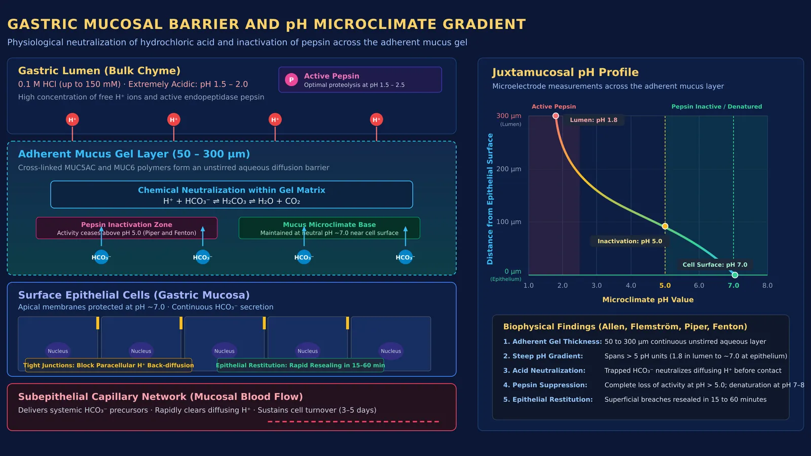
According to measurements compiled by Allen and Flemström, this adherent mucus gel layer maintains a thickness between 50 and 300 µm in the human stomach. Crucially, the gel does not act merely as a physical barrier. Surface epithelial cells actively secrete bicarbonate ions (HCO3⁻) into the base of the gel through apical anion exchangers. Because the gel forms a stable, unstirred aqueous layer, it prevents convection and slows the diffusion of luminal hydrogen ions (H⁺) toward the tissue.
As hydrogen ions diffuse downward from the lumen, they encounter ascending bicarbonate ions trapped within the gel matrix. The neutralization reaction (H⁺ + HCO3⁻ ⇌ H2CO3 ⇌ H2O + CO2) eliminates the acid before it reaches the epithelial surface. This establishes a steep microclimate gradient across less than a millimetre: while the lumen churns at pH 1.5 to 2.0, the juxtamucosal microclimate at the apical cell membranes remains at a neutral pH near 7.0.
When studying biophysical transport and chemical equilibria across biological membranes, visualizing unstirred layers can feel abstract. The Fermi Concept Library covers every concept in physics, chemistry, biology, mathematics and computing, each with its own lesson and its place in the map.
Why does active pepsin not break down stomach cells?
Neutralizing hydrochloric acid solves only half the challenge. Gastric juice also contains pepsin, which cleaves peptide bonds in dietary proteins and can damage cellular proteins if left unchecked. Research on pepsin activity and stability curves by D. W. Piper and B. H. Fenton reveals the physiological mechanisms that prevent tissue destruction:
Inactive zymogen secretion: Chief cells in gastric glands produce pepsin as an inactive precursor called pepsinogen. An inhibitory activation peptide masks its catalytic cleft, preventing intracellular proteolysis during storage and secretion.
Luminal acid activation: Pepsinogen is activated only after entering the acidic lumen below pH 5.0, where low pH triggers conformational unfolding and peptide cleavage to generate active pepsin.
Inactivation at the neutral boundary: Piper and Fenton demonstrated that human pepsin exhibits peak activity between pH 1.5 and 2.5, but becomes inactive above pH 5.0 and undergoes complete, irreversible inactivation as pH approaches 8.0.
Because the mucus gel maintains a juxtamucosal microclimate near pH 7.0, any pepsin entering the mucus layer is catalytically inactive before it can contact apical cell membranes. In addition, the cross-linked mucin matrix acts as a steric filter that prevents larger enzyme molecules from penetrating the gel. For students reviewing enzyme kinetics and denaturation curves, our guide to enzymes for NEET biology outlines essential reaction principles.
How do epithelial cells repair damage so quickly?
Mechanical abrasion from food churning and occasional acid penetration inevitably damage surface cells. The mucosa protects and repairs itself through two cellular mechanisms.
Adjacent surface epithelial cells are joined by apical tight junctions composed of claudin and occludin proteins. As reviewed by Tyler J. Caron and colleagues, these tight junctions provide high electrical resistance across the epithelium and prevent paracellular back-diffusion of luminal acid into subepithelial tissue.
When superficial cells are injured, the mucosa triggers an ultra-rapid repair mechanism called epithelial restitution. In their foundational study in the Annual Review of Physiology, William Silen and Susumu Ito showed that superficial epithelial breaches are resealed within 15 to 60 minutes. During restitution, surviving cells in adjacent gastric pits flatten, extend lamellipodia, and migrate across the intact basement membrane to restore continuity without waiting for cell division.
Beneath the basement membrane, rich microvascular blood flow removes back-diffusing protons while delivering oxygen and bicarbonate precursors. In addition, adult stem cells in the gastric neck region divide continuously, renewing the entire surface epithelial lining every 3 to 5 days, as noted in mucosal turnover studies cited by Caron and colleagues.
How should students approach mucosal barrier exam questions?
Questions on gastric secretions, enzyme activation, and mucosal protection appear regularly in CBSE Class 11 biology and NEET-UG. Synthesizing the benchmarks established by Allen and Flemström and Silen and Ito gives students an empirical framework to solve Assertion-Reason questions. The NCERT biology textbook specifically highlights that mucus and bicarbonates protect the mucosal epithelium from excoriation by concentrated hydrochloric acid.
When evaluating exam questions, verify each tier of the mucosal barrier in sequence:
Secretory tier (precursor safety): Chief cells produce pepsinogen as an inactive zymogen, preventing autodigestion of intracellular structures prior to luminal release.
Pre-epithelial tier (chemical buffer): Surface mucous cells maintain an adherent mucus gel of 50 to 300 µm that traps secreted bicarbonate, sustaining a neutral microclimate near pH 7.0 against luminal acid of pH 1.5 to 2.0.
Epithelial tier (cellular barrier): Apical tight junctions block paracellular proton diffusion, while epithelial restitution reseals superficial lesions within 15 to 60 minutes.
Subepithelial tier (vascular clearance): Robust mucosal blood flow sweeps away penetrating acid and replenishes bicarbonate precursors.
To master the exact terminology tested in exams, review our guide on mastering NCERT lines for NEET biology. For challenging multi-step physiology problems, the Fermi Problem Library provides worked problems with a step-by-step solver that explains the reasoning rather than printing an answer. When visualizing how concentration gradients form across mucosal layers, the Fermi Simulation Lab provides interactive simulations for the ideas that are easier to see than to read.


