
On this page
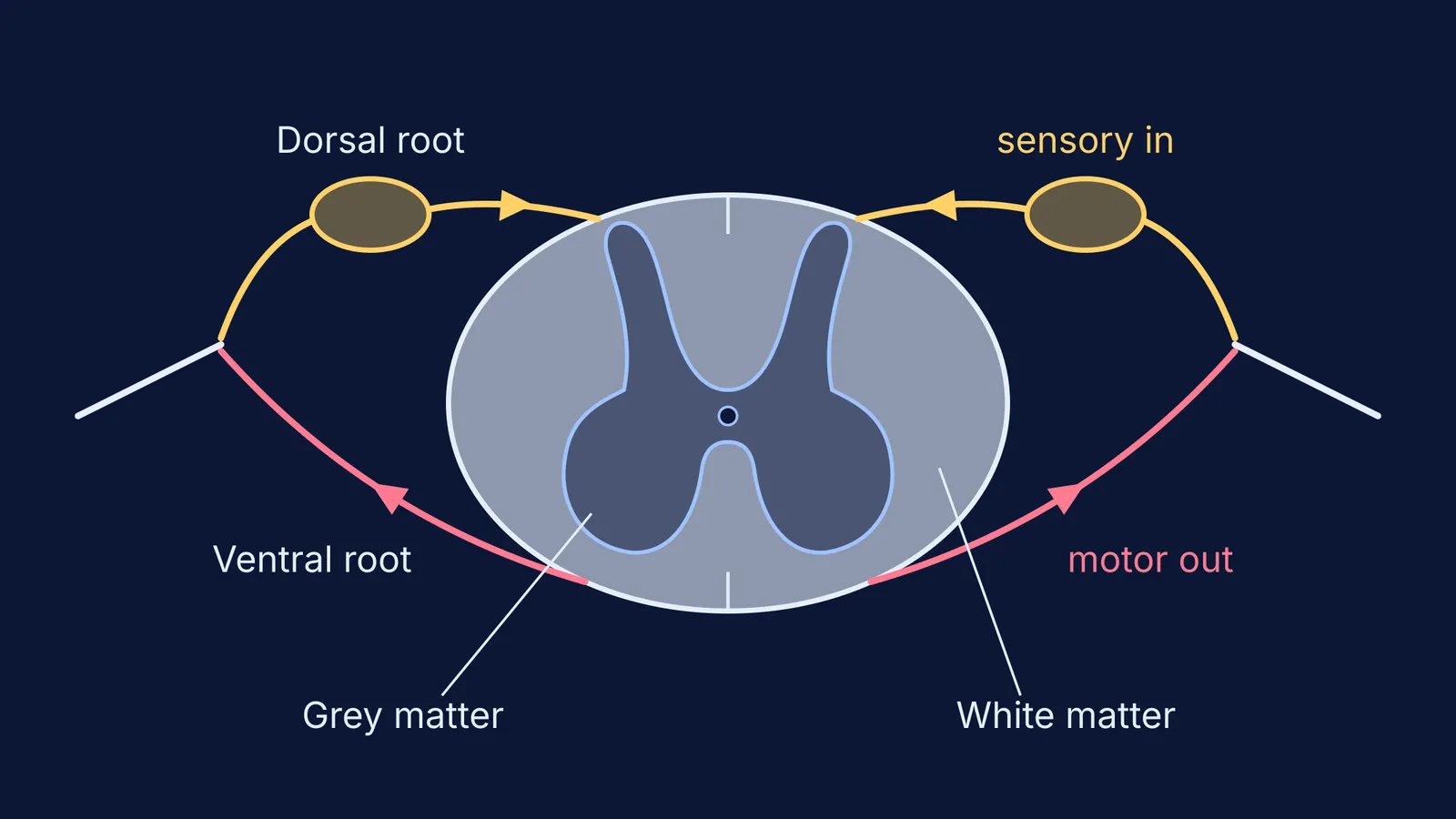
Cut a spinal cord across and you see a grey butterfly inside a pale oval. Cut the cerebrum and the order flips: a grey rind outside, pale tissue beneath. The difference comes from how each part develops.
What are grey matter and white matter in the spinal cord?
Grey matter is mostly neuron cell bodies, and white matter is mostly bundles of nerve fibres. The StatPearls review Neuroanatomy, Spinal Cord Morphology describes the cord’s grey matter as “a collection of cell bodies” with “an H-shaped or butterfly pattern” in cross-section, surrounded by white matter made of fibre tracts. The same review says the adult cord is up to 45 cm long in men and 43 cm in women, ending at the level of the L1 to L2 vertebrae.
The OpenStax chapter on the central nervous system names the parts of the butterfly:
Posterior (dorsal) horn: sensory processing.
Anterior (ventral) horn: sends motor signals to skeletal muscles.
Lateral horn: autonomic motor neuron cell bodies, found only in the thoracic, upper lumbar and sacral regions.
The white matter is split into posterior, anterior and lateral columns, where ascending tracts carry sensory information up to the brain and descending tracts carry motor commands down, as the same OpenStax chapter explains. White matter is white because its axons are insulated by myelin, a lipid-rich substance, according to OpenStax on the structure of the nervous system. Our explainer on myelinated and non-myelinated nerve fibres covers what that sheath does.
Why is grey matter inside the spinal cord but outside in the brain?
The answer lies in how each part grows from the embryo’s neural tube. The University of North Carolina’s embryology notes on the spinal cord describe layers in the tube wall, including “a mantle layer of differentiating neurons that will form the gray matter of the spinal cord, and a marginal layer that contains nerve fibers and will be the white matter.”
In the spinal cord, neurons stay close to the central canal and their fibres gather around the outside. So white matter ends up outside and grey matter stays in.
The cerebrum adds a step. Its neurons are born beside the fluid-filled ventricle and then travel outward. A 2016 review by Hatanaka and colleagues in the Proceedings of the Japan Academy, Series B notes that newborn neurons “migrate radially from the ventricular zone” along guiding glial fibres. They settle near the surface as the cerebral cortex, which OpenStax calls “a continuous layer of gray matter” covering the cerebrum. The fibre tracts are left underneath, so in the cerebrum the white matter sits deep.
The flip is not complete: the OpenStax chapter also describes basal nuclei, clusters of grey matter located deep in the cerebrum.
What do the dorsal and ventral roots carry?
Each spinal nerve joins the cord through two roots. OpenStax’s chapter on the peripheral nervous system states that “the sensory axons enter the spinal cord as the dorsal nerve root” and “the motor fibers, both somatic and autonomic, emerge as the ventral nerve root.”
OpenStax counts 31 spinal nerves, each named for the cord level where it emerges, and every one is mixed: sensory and motor axons travel together, then separate into the two roots. The swelling on the dorsal root is the dorsal root ganglion, which StatPearls describes as holding the cell bodies of the sensory pathway.
So the roots line up with the horns: sensory fibres come in at the back, towards the dorsal horn, and motor fibres leave from the ventral horn at the front. Join the two and you have the pathway of a reflex arc, from receptor to effector.
How do you answer root-damage questions?
When a question asks what happens if one root is cut, reason it out:
Name what the root carries: dorsal is sensory and goes in, ventral is motor and goes out.
Find what is lost: a dorsal root cut takes away feeling; a ventral root cut takes away the muscle’s ability to move.
Check the reflex: a reflex needs both the route in and the route out, so cutting either root stops it.
Worked example. Suppose the dorsal root at one level is cut. The area it serves loses sensation, because sensory signals cannot reach the cord. The muscles can still move on a command from the brain, because the ventral root is intact. The reflex at that level is lost, because the input side of the arc has gone.
Swap it for a ventral root cut and the answer reverses: feeling stays, while voluntary movement and the reflex go.
If a term like ganglion or tract is still fuzzy, the Fermi Concept Library gives each biology concept its own lesson and shows its place in the map. For practice, the Fermi Problem Library has worked problems with a step-by-step solver that explains the reasoning rather than printing an answer.
For where the cord fits in the whole system, read our guide to nervous system divisions: CNS, PNS, somatic and autonomic.