On this page
You will meet this comparison in Class 11 biology and again in NEET-UG-style questions, usually as a two-column answer. The facts are short. What examiners test is whether you know why myelin matters, and that in peripheral nerves "non-myelinated" does not mean "no Schwann cells".
What is the difference between myelinated and non-myelinated nerve fibres?
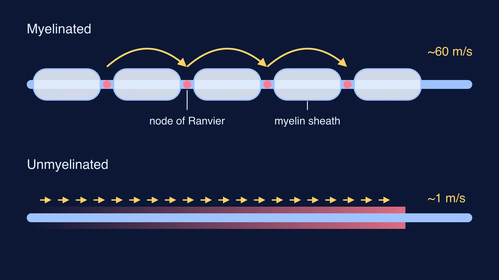
A myelinated fibre is an axon wrapped in myelin, a lipid-rich sheath that, in the words of OpenStax Anatomy and Physiology, "facilitates the transmission of electrical signals along the axon". The wrapping is not continuous: it breaks at regular gaps called nodes of Ranvier.
A non-myelinated (unmyelinated) fibre has no such sheath, so the impulse has to travel along every bit of membrane. The difference shows up as speed.
Unmyelinated C fibres conduct at about 1 m/s, and thinly myelinated Aδ fibres at about 10 m/s, according to a 2014 review in Practical Neurology.
If you still mix up "nerve", "neuron" and "nerve fibre", sort that out first with our explainer on nerve vs neuron vs nerve fibre.
What do Schwann cells and nodes of Ranvier do?
Myelin is made by glial cells, and which cell depends on where you are. In the peripheral nervous system, Schwann cells make it: OpenStax notes that "a Schwann cell wraps around a portion of only one axon segment and no others". In the brain and spinal cord, oligodendrocytes do the job, and one oligodendrocyte reaches out to several axon segments.
So along one peripheral axon you get a row of Schwann cells, each covering its own segment, with a node of Ranvier between neighbours. At each node the bare axon membrane is exposed, and that is where the impulse is regenerated.
That description of Remak bundles comes from the Practical Neurology review and a 2023 review of neuron and Schwann cell interactions.
How does saltatory conduction make impulses faster?
In an unmyelinated axon, voltage-gated sodium channels open one patch after the next along the whole membrane. OpenStax calls this continuous conduction and explains that it is slow because channels keep opening all the way down the axon.
In a myelinated axon, the action potential is regenerated only at the nodes, so it effectively leaps from one node to the next. That is saltatory conduction, from the Latin saltare, "to leap" (OpenStax). Diameter matters too: depolarisation spreads faster down a wide axon than a narrow one, so thick myelinated fibres are the quickest of all.
Worked example: how long does the impulse take?
Use time = distance ÷ speed, and imagine the same 1 m path for each fibre type.
Myelinated motor nerve: a study of 290 participants aged 17 to 21 in Punjab measured median motor nerve conduction at about 60 m/s. Time = 1 ÷ 60 ≈ 0.017 s, or about 17 ms.
Thinly myelinated Aδ fibre at about 10 m/s (Practical Neurology): 1 ÷ 10 = 0.1 s.
Unmyelinated C fibre at about 1 m/s: 1 ÷ 1 = 1 s.
Same distance, roughly sixty times longer for the slowest fibre. Keep everything in metres and seconds and the arithmetic stays clean. Our post on predicting conduction speed in myelinated and unmyelinated nerves takes this idea further.
Where are myelinated and non-myelinated fibres found?
Brain and spinal cord: white matter is myelinated. It "is white because axons are insulated by a lipid-rich substance called myelin" (OpenStax).
Peripheral nerves: myelinated axons sit alongside Remak bundles of unmyelinated C fibres (Practical Neurology).
Pain and temperature: thinly myelinated Aδ fibres carry cold and pain signals, while unmyelinated C fibres carry warmth and noxious mechanical, thermal and chemical input (same review).
Autonomic nervous system: thin myelinated fibres contribute to preganglionic fibres and C fibres to postganglionic fibres, which supply sweat glands, blood vessels and the heart (same review).
Your textbook may phrase the locations in its own way. Learn its exact wording for board answers, and use the list above to understand why.
How do you write this comparison in an exam?
A five-point answer covers most of what is asked:
Sheath: myelin present vs absent.
Cells: Schwann cells (peripheral) or oligodendrocytes (brain and spinal cord) vs, in peripheral nerves, non-myelinating Schwann cells that wrap no myelin.
Nodes of Ranvier: present vs absent.
Conduction: saltatory, node to node, vs continuous along the membrane.
Speed and location: fast, in white matter and peripheral nerves, vs slow, in C fibres for warmth and pain and in postganglionic autonomic fibres.
Add a quick sketch if marks allow. Our guide on how to draw and label a neuron diagram walks through the Schwann cell, node and myelin labels.
If myelin still feels like a fact without a place, the Fermi Concept Library gives every concept in biology its own lesson and its place in the map, so you can see where it sits among the ideas around it.
Tonight, close the book and write the five-point comparison from memory, then check it against this page.