

On this page
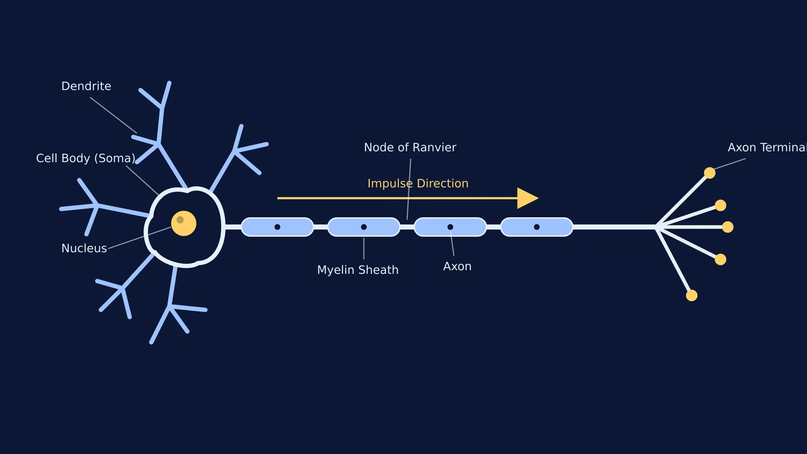
Drawing a neuron (nerve cell) is one of the most reliable questions in board and competitive biology exams. More than just a test of artistic skill, the diagram shows how the nervous system receives and transmits signals across the human body.
While it is tempting to just stare at the textbook and hope the image sticks, research shows you learn faster with a pencil in your hand. Students who drew pictures of concepts recalled nearly double the amount of information compared to those who wrote them out, according to a 2016 study by researchers at the University of Waterloo published in the Quarterly Journal of Experimental Psychology.
Here is a practical, step-by-step guide to sketching the standard multipolar neuron, understanding its function, and getting the labels exactly right for your exams.
Why does drawing beat just looking?
When you read text or passively scan a diagram, your brain only encodes the information visually and semantically. Drawing forces you to use your motor skills to translate a three-dimensional biological structure onto a two-dimensional page.
This process of "generative learning" integrates multiple neural pathways, making the memory trace far stronger. The physical act of drawing clarifies the relationships between different parts of the cell, so you actually understand how the structures work together rather than just memorizing their names. According to the researchers behind the study, the drawing effect works because it combines semantic meaning, visual imagery, and physical motor action into one rich memory trace.
How to draw a neuron diagram step-by-step?
You do not need to be an artist to score full marks on this question; the goal is structural accuracy and clarity. Follow these steps to build the cell from the center out:
Cell Body (Soma): Start on the left side of your page. Draw a star-like or irregular circular shape. This is the main body of the cell where metabolic processes happen.
Nucleus: Place a solid, dark circle right in the middle of the cell body.
Dendrites: From the edges of the cell body, draw short, tree-like branches that split into smaller twigs. These should radiate outward in all directions.
Axon: Choose one side of the cell body and draw a single, long, continuous tube extending to the right. It should be significantly longer than any of the dendrites.
Myelin Sheath: Along the length of the axon, draw a series of rounded rectangular blocks, like beads on a string. This represents the protective fat layer.
Nodes of Ranvier: Leave small, distinct gaps between the myelin sheath blocks so the bare axon is visible.
Axon Terminals (Nerve Endings): At the far right end of the axon, draw it splitting into several tiny branches, each ending in a small bulb (the synaptic knob).
What are the essential neuron labels?
A diagram is incomplete without correct, straight labeling lines. Keep your labels aligned to one side of the page if possible, and include these core parts along with a clear understanding of their functions:
Dendrites: The receiving branches that pick up incoming nerve impulses from other cells and carry them toward the cell body.
Cell Body (Soma): The metabolic center of the neuron that processes incoming signals and keeps the cell alive.
Nucleus: The central organelle containing the genetic material that controls cell activity.
Axon: The long conductive fiber that carries electrical impulses away from the cell body toward the next cell.
Myelin Sheath: The fatty insulating layer around the axon that greatly increases the speed of signal transmission.
Nodes of Ranvier: The small gaps in the myelin sheath where the electrical impulse jumps from node to node to travel faster via saltatory conduction.
Axon Terminals: The nerve endings where chemical neurotransmitters are released across the synapse to communicate with the next cell.
Understanding these structures in detail helps you make sense of much larger nervous system topics, such as control and coordination in Class 10: reflexes, brain and hormones.
How do you check your neuron diagram for mistakes?
Before you move on to the next exam question, check your drawing against this practical framework to ensure you have not missed any easily avoidable mistakes:
Is the nucleus drawn clearly inside the cell body?
Are the dendrites visibly much shorter and more branched than the single long axon?
Did you leave clear, visible gaps (Nodes of Ranvier) between the myelin blocks?
Do the axon terminals end in distinct bulbs rather than sharp points?
Is the direction of the impulse marked with an arrow traveling away from the cell body?
Building biology concepts from the ground up
If you find yourself stuck trying to link microscopic cell structure to function in the nervous system—or wondering how a single neuron connects to the parts of the human brain: forebrain, midbrain and hindbrain roles—a static textbook is not always enough.
The Fermi Concept Library contains every concept in physics, chemistry, biology, mathematics and computing, each with its own lesson and its place in the map. When you bring a tough biology topic, Fermi's AI tutor works out what you already know, builds a plan for the session, and teaches through it—drawing the figure and checking understanding as it goes.


