
On this page
Your skin is an organ, not just a wrapper. The OpenStax Anatomy and Physiology textbook describes it as tissues that “work together as a single structure” (OpenStax, Layers of the Skin). For school biology you need three things: the layers, what sits inside them, and one neat labelled section.
In the 2026-27 ICSE syllabus, as reproduced by the publisher Oswaal, “Structure and functions of skin” is a Class 9 topic. It covers the parts of the skin, its glands, hair and nails, and heat regulation by vasodilation and vasoconstriction (ICSE Class 9 Biology syllabus 2026-27).
What are the layers of human skin?
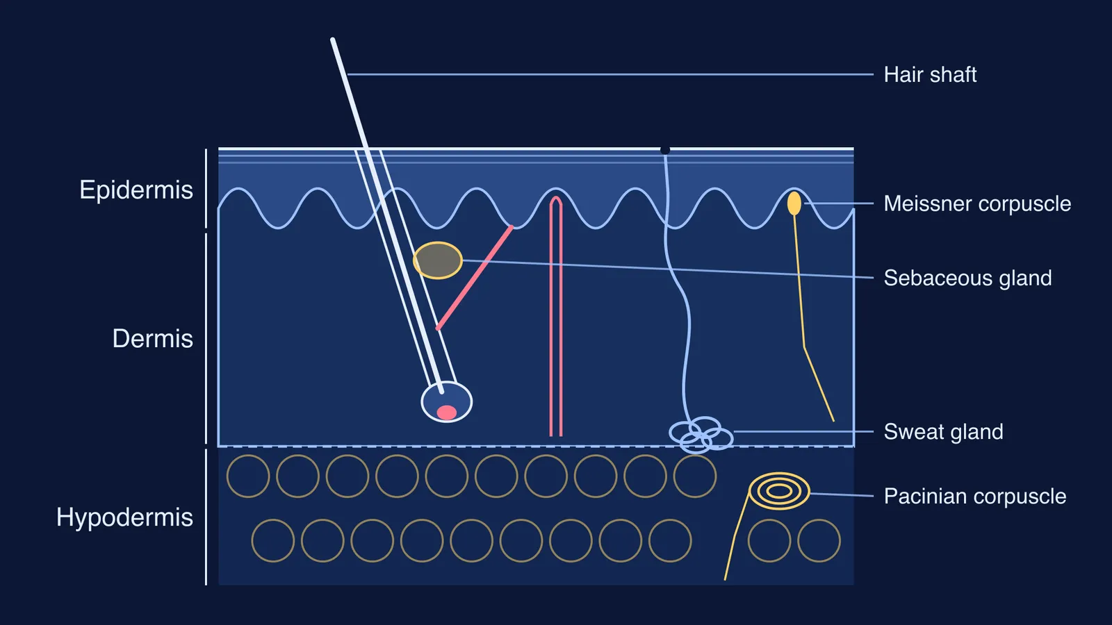
Skin has three layers, from the outside in: epidermis, dermis and hypodermis (OpenStax).
Epidermis: the outer layer. It has no blood vessels (it is avascular). From deep to surface its sub-layers are the stratum basale, stratum spinosum, stratum granulosum and stratum corneum. Thick skin on the palms, soles and digits adds a fifth, the stratum lucidum, between the granulosum and the corneum.
Dermis: two layers of connective tissue, the papillary layer on top and the thicker reticular layer below.
Hypodermis: loose areolar connective tissue and adipose (fat) tissue that cushions the body and anchors the skin.
The main cells of the epidermis are keratinocytes, which make the protein keratin, and melanocytes, which make the pigment melanin. The whole epidermis is replaced in about 4 weeks (OpenStax). The surface cells are dead and full of keratin, the same reason cutting your hair and nails does not hurt.
Where are the sweat glands, oil glands and hair follicles?
All three grow from the epidermis and reach down into the dermis, sometimes as far as the hypodermis (OpenStax, Accessory Structures of the Skin).
Hair follicle: a pocket of epidermis pushed down into the dermis. At its base is the hair bulb, wrapped around the hair papilla, which carries capillaries and nerve endings.
Arrector pili: a small smooth muscle joined to each hair root. When it contracts, the hair stands up.
Sebaceous (oil) glands: they release sebum, a mix of lipids that lubricates and waterproofs skin and hair.
Sweat glands: eccrine glands are coiled glands deep in the dermis, found all over the body and especially on the palms, soles and forehead. Apocrine glands sit with hair follicles in hairy areas such as the armpits.
A 2019 review by Lindsay Baker in the journal Temperature puts the total at about 2 to 4 million eccrine sweat glands, with about 250 to 550 glands per square centimetre on the palms and soles (Baker 2019).
The dermis also carries touch receptors. Meissner corpuscles sit in the dermal papillae and sense light touch; Pacinian corpuscles lie deep in the dermis or in the tissue below it and sense vibration and deep pressure (OpenStax, Functions of the Integumentary System; OpenStax, Sensory Perception).
How does skin control body temperature?
When you are hot, arterioles in the dermis widen (vasodilation) so blood can lose heat through the skin, and an active person can make as much as 0.7 to 1.5 litres of sweat per hour (OpenStax). When you are cold, the arterioles narrow (vasoconstriction) to keep heat in.
School books also list skin as an excretory organ; see the role of lungs, liver and skin in excretion. Keep it in proportion: Baker's review found that sweat's role in removing waste seems minor compared with the kidneys and gut (Baker 2019).
How do you draw the labelled skin section?
Build the drawing in this order:
Draw three bands. Keep the epidermis thin, the dermis widest and the hypodermis at the bottom.
Make the lower edge of the epidermis wavy. The bumps are the dermal papillae.
Draw a slanted hair follicle into the dermis, with a round bulb and papilla at the bottom and the hair shaft coming out.
Add a sebaceous gland beside the follicle and the arrector pili running from the follicle up to the underside of the epidermis.
Draw a coiled sweat gland deep in the dermis, with a duct running up to a pore on the surface.
Put a capillary loop into one papilla, a Meissner corpuscle in another, and a layered Pacinian corpuscle deep down.
Fill the hypodermis with round fat cells.
Label on one side, with straight lines that never cross.
To test yourself, cover your notes, draw the section from memory, then tick off nine labels: hair shaft, sebaceous gland, arrector pili, sweat gland, sweat pore, blood capillary, Meissner corpuscle, Pacinian corpuscle, fat cells.
If one part keeps going blank, the Fermi Concept Library has a lesson for each biology concept and shows where it fits in the map. Fermi's AI tutor can draw the figure with you and check your understanding as it goes.
Next, try the same method on the labelled diagram of the human heart for Class 10.