On this page
Clipping your fingernails or getting a haircut is completely painless, yet plucking a single hair or slipping a clipper into the skin beneath your nail triggers sharp, immediate pain. The reason lies in the boundary between dead structures and living tissue.
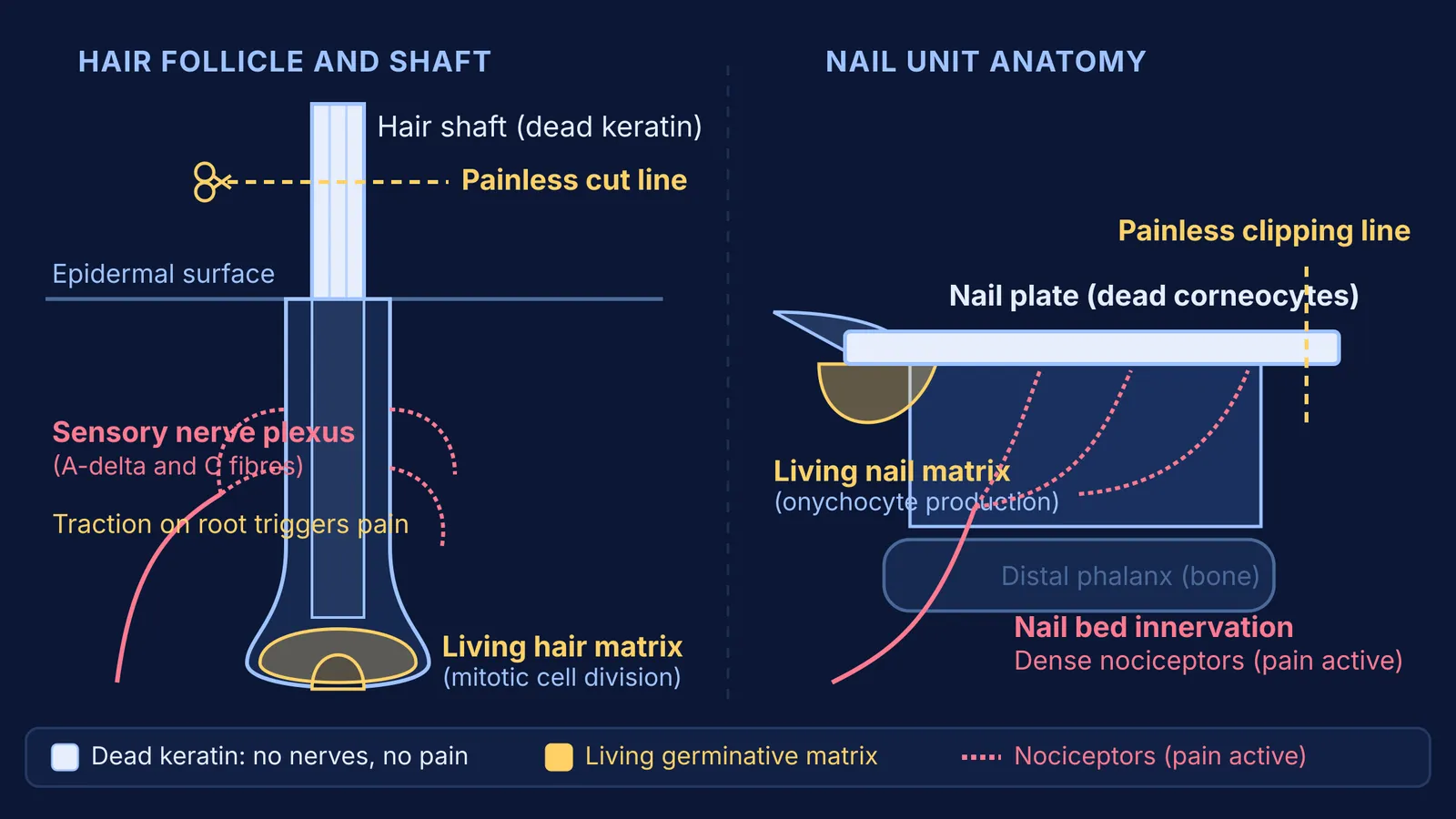
The hair shaft extending past your scalp and the nail plate over your fingertips consist entirely of dead, cornified cells. According to StatPearls Hair Anatomy, the hair shaft contains no sensory nerve endings, no pain receptors, no blood supply, and no living cell membranes capable of generating action potentials.
Sensory nerves register pain only when stimuli depolarize living membranes, activating high-threshold neurons called nociceptors. In an exposed hair strand or the distal edge of a nail, no living cells exist to depolarize. Trimming them is biologically equivalent to cutting a thread of wool or dry paper.
How do living cells turn into dead keratin structures?
Every hair and nail originates in a living germinative zone beneath the skin: the hair matrix at the follicle bulb and the nail matrix under the proximal nail fold. Here, basal keratinocytes divide rapidly, pushing older cells outward into a pathway called cornification. As documented by Leopold Eckhart and colleagues in Biochimica et Biophysica Acta, cornification is a specialized programmed cell death that dismantles internal organelles while preserving a durable protein framework.
During this differentiation, nucleases degrade nuclear DNA, and organelles are cleared. Research in Apoptosis by Karin Jaeger and colleagues showed that nail keratinocytes rely on autophagy for the bulk degradation of intracellular enzymes and protein complexes, while selectively sparing the structural keratin cytoskeleton and cell-adhesion desmosomes.
As metabolic activity ceases, cells pack with hard alpha-keratin. As outlined in our guide to protein functions and structural proteins, hard alpha-keratins are rich in cysteine residues forming covalent disulfide bonds (-S-S-). Transglutaminase enzymes cross-link an insoluble cornified envelope beneath the plasma membrane, locking dead cells into a rigid, waterproof structure.
When mapping cell differentiation or clarifying how epithelial tissues assemble, the Fermi Concept Library covers every concept in biology with its own lesson and its place in the topic map.
Why does pulling hair or cutting the nail bed hurt so much?
If hair shafts and nail plates are completely inert, why is pulling hair painful, and why does cutting too close to the nail bed hurt?
Both structures are anchored in sensitive zones of the peripheral nervous system. According to StatPearls Hair Anatomy, each hair follicle root is wrapped by a peritrichial nerve plexus of longitudinal A-delta fibers and circumferential C-fibers. Healthy scalp hair follicles, numbering between 80,000 and 120,000 terminal hairs on an adult head according to StatPearls Hair Anatomy, grow at an average rate of 0.35 mm per day. Scissors slice through inert keratin without disturbing the dermis. But pulling hair turns the shaft into a mechanical lever that transmits tensile force down into the follicle, stretching connective tissue and firing nociceptors.
A similar arrangement protects the digits. According to StatPearls Nail Anatomy, while fingernails grow at roughly 3 mm per month and toenails at approximately 1 mm per month, the underlying nail bed is rich in digital sensory nerves. Trimming the distal white edge cuts only dead onychocytes. Cutting into the hyponychium or vascular bed crushes living tissue, releasing chemicals that stimulate nociceptors. You can explore how these sensory signals travel in our guide to conduction speeds of myelinated and unmyelinated nerve fibres.
Just as epidermal keratinocytes synthesize protective barriers while melanocytes distribute pigment granules, as discussed in our article on how epidermal cells respond to sunlight and produce melanin, hair and nails function as shields that protect sensitive underlying nerve networks from mechanical trauma.
When working through physiology problems or reviewing structural tissues, the Fermi Problem Library offers worked problems with a step-by-step solver that explains the reasoning rather than printing an answer.
What biological principles distinguish painless cuts from pain?
Four structural distinctions explain the difference between painless trimming and severe pain:
Distal free edge versus vascular bed: The white distal crescent of the nail plate has completely separated from the vascular nail bed and consists solely of dead, cornified onychocytes. Trimming remains painless as long as cutting edges stay within this insensitive zone.
Protective hyponychium junction: The tissue junction beneath the distal nail plate forms a living, innervated anatomical seal. Breaching this junction excites nociceptive free nerve endings and breaks the physical barrier against foreign microbes.
Hard keratin versus epidermal desquamation: Surface skin sheds flexible soft keratin during desquamation, whereas hair shafts and nail plates consist of permanent hard alpha-keratin reinforced by disulfide cross-links that resist mechanical degradation.
Lever action on follicle roots: Scissors shear inert keratin without disturbing dermal nerves. In contrast, pulling hair exerts tensile force along the shaft, mechanically distorting the follicle root and firing the peritrichial nerve plexus.