On this page
If you are drawing the heart for a Class 10 Biology answer, the version to learn is the one in your NCERT textbook: a schematic cut-through view, not a realistic organ. Here is what goes on it, why one side is blue, and how to draw it neatly.
What should a labelled diagram of the human heart show?
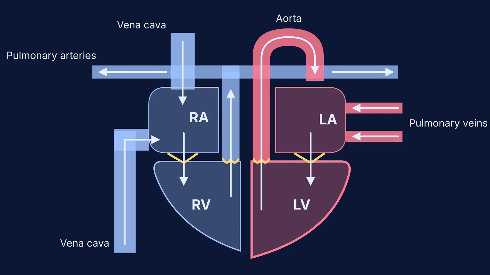
The NCERT Class 10 Science chapter Life Processes calls its heart figure a "schematic sectional view of the human heart" (Figure 5.10), and it labels ten parts (NCERT, Life Processes, 2026-27 reprint):
Right atrium and left atrium, the two upper chambers
Right ventricle and left ventricle, the two lower chambers
Septum, the dividing wall between the right and left sides
Vena cava from the upper body and vena cava from the lower body
Pulmonary arteries and pulmonary veins
Aorta
The same chapter explains the logic. The heart is a muscular organ about as big as your fist, with separate chambers so that oxygen-rich blood does not mix with blood carrying carbon dioxide. Ventricles have thicker muscular walls than atria because they pump blood out to the organs, and valves stop blood flowing backwards when the chambers contract (NCERT).
The NCERT figure does not name the valves. If your teacher asks for them, there are four: the tricuspid valve between the right atrium and right ventricle, the pulmonary semilunar valve where blood leaves the right ventricle, the mitral (bicuspid) valve between the left atrium and left ventricle, and the aortic semilunar valve at the base of the aorta (OpenStax Anatomy and Physiology 2e, section 19.1). Our guide to the names and positions of the heart valves goes further.
Why is the right side of a heart diagram coloured blue?
Blue marks deoxygenated blood and red marks oxygenated blood. It is a labelling convention, not the real colour: deoxygenated blood is darker but still red (Medical News Today, Is blood blue?).
The right side of the heart collects blood from the body and sends it to the lungs, so it is shaded blue. The left side receives oxygen-rich blood from the lungs and pumps it to the body, so it is shaded red. NCERT traces both routes: lungs to left atrium to left ventricle to body, and body to right atrium to right ventricle to lungs (NCERT).
Two vessels break the "arteries are red" habit. The pulmonary trunk carries deoxygenated blood from the right ventricle and splits into the pulmonary arteries to the lungs, while the pulmonary veins carry highly oxygenated blood into the left atrium (OpenStax). So the pulmonary arteries are blue and the pulmonary veins are red.
How do you draw a neat labelled heart diagram step by step?
Work in pencil first, in this order:
Draw a rounded outline, broad at the top and narrowing to a blunt point that leans to your right.
Draw the septum down the middle, then a cross line about a third of the way down. The atria should be smaller than the ventricles.
Thicken the ventricle walls, making the left ventricle (on your right) the thickest (OpenStax).
Add the vessels: vena cava into the right atrium from above and below, pulmonary artery out of the right ventricle, pulmonary veins into the left atrium, aorta arching out of the left ventricle.
If valves are asked for, draw small flaps between each atrium and ventricle and at the bases of the pulmonary artery and aorta.
Add flow arrows, then colour the right side blue and the left side red.
Label on the outside with straight, horizontal pointer lines, and write the figure title underneath.
Then check your arrows against this trace: vena cava, right atrium, tricuspid valve, right ventricle, pulmonary valve, pulmonary artery, lungs, pulmonary veins, left atrium, mitral valve, left ventricle, aortic valve, aorta, body. Blood goes through the heart twice in each cycle round the body, which NCERT calls double circulation (NCERT); our explainer on double circulation in humans walks through it.
If double circulation still feels like a list rather than a loop, the Fermi Concept Library has a lesson for every biology concept, each with its place in the map, so you can see what the heart connects to.
Which mistakes should you check for in a heart diagram?
Before you hand in, check for these:
Right and left swapped, so the blue side ends up on your right
Pulmonary artery coloured red, or pulmonary veins coloured blue
Atria as big as the ventricles, or every wall the same thickness
Arrows missing, or pointing backwards through a valve
Labels crossing each other or crammed inside chambers
No figure title
The same habits carry over to the nephron, the alimentary canal and the eye; see our list of diagrams to practise for CBSE Class 10 Science. If you want your understanding checked as you go, Fermi's AI tutor draws the figure and decides whether to explain, ask or show, so "why is the left ventricle thickest?" gets reasoning, not just a label.