
On this page
If you learnt mitosis from the animal-cell diagram in NCERT, a plant cell looks like it is missing a part. The textbook says the centrosome moves to the poles and "radiates out microtubules called asters", and that the asters plus spindle fibres form the mitotic apparatus (NCERT Class 11 Biology, Chapter 10). Land-plant cells have no centrosome, yet they still build a spindle.
What is the difference between astral and anastral mitosis?
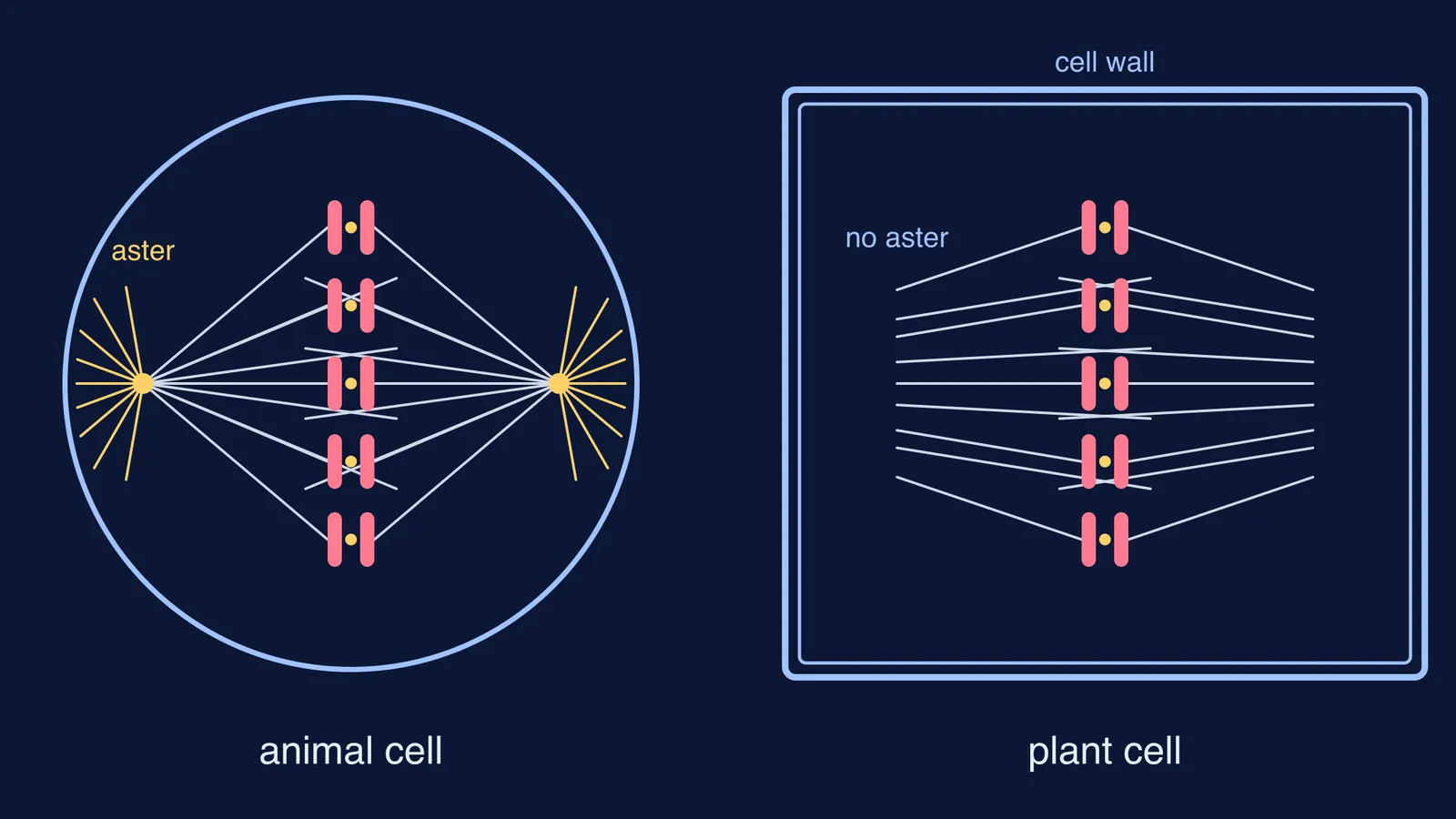
Astral mitosis is division with asters: star-shaped rays of microtubules around each centrosome at the spindle poles, as drawn in the NCERT animal-cell figure (NCERT, Chapter 10). Anastral mitosis is division without asters, and it is the usual kind in flowering plants.
Land plants lack centrosomes, which are the main microtubule organising centre in animal body cells, according to a review by Moé Yamada and Gohta Goshima in the journal Biology (Yamada and Goshima, 2017). Liverworts are a partial exception: the review describes short-lived polar organisers, with no centriole, that make astral microtubules in prophase.
The review searched for more than 100 animal spindle assembly factors and found them largely conserved in plants, except centrosome components and the cytoplasmic dynein motor complex (Yamada and Goshima, 2017). The visible result is the shape: the review describes the plant metaphase spindle as generally barrel-shaped rather than fusiform, because each pole is not tightly focused at one point.
If centrosome, centriole and centromere still blur together for you, sort them first with our post on centromere vs centrosome vs centriole.
Where does the spindle come from in plant cells?
A preprophase band sets the plan. Before mitosis, a transient ring of microtubules and actin filaments forms just under the membrane of plant cells and marks the future division plane (Li, Sun and Ren, 2015); it generally breaks down by the end of prophase (Yamada and Goshima, 2017).
The nuclear envelope acts as the starting surface. In plant cells the spindle originates from the nuclear envelope in prophase (Li, Sun and Ren, 2015). In most flowering-plant cells, microtubules collect around the nucleus as two "polar caps", also called the prophase spindle (Yamada and Goshima, 2017).
New microtubules grow from old ones and near chromosomes. After the nuclear envelope breaks down, more microtubules branch off existing ones (a process that depends on a protein complex called augmin) and form close to the chromosomes (Yamada and Goshima, 2017).
How does cytokinesis differ in plant and animal cells?
In an animal cell, a furrow appears in the plasma membrane and deepens until it pinches the cell in two (NCERT, Chapter 10).
A plant cell cannot pinch, because it sits inside a relatively inextensible cell wall. Instead, wall formation starts in the centre of the cell and grows outward to meet the side walls, beginning with a cell plate that becomes the middle lamella (NCERT, Chapter 10).
The cell plate grows inside the phragmoplast, a plant-specific structure of microtubules and actin filaments that guides vesicles to build the new wall (Li, Sun and Ren, 2015). It forms in telophase and expands outward towards the cell edge (Yamada and Goshima, 2017). Animals divide from the outside in, plants from the inside out. Our guide to cytokinesis in plant and animal cells has the full comparison.
How do you draw animal and plant metaphase side by side?
Use the same chromosome layout in both sketches so the examiner sees that only the spindle and the boundary change.
Outline: animal cell round with a single membrane; plant cell box-shaped with a thick cell wall drawn as a double line.
Poles: animal cell has a dot (centrosome) at each pole with short rays (aster) pointing away from the spindle; plant cell has no dot and no rays, just a broad, flat pole.
Spindle shape: animal fibres converge to a point (spindle-shaped, fusiform); plant fibres converge only partly towards broad poles (barrel-shaped).
Chromosomes: in both, each chromosome sits on the metaphase plate with one sister chromatid facing each pole and a fibre attached at each kinetochore.
Labels: centrosome and aster on the animal cell only; cell wall on the plant cell only; spindle fibre, kinetochore and metaphase plate on both. Write "no centrioles, anastral" beside the plant sketch.
To practise naming the stage from a figure, work through how to identify mitosis and meiosis stages from a diagram.
How should you write a 3-mark answer?
One fact per line, and remember that "no centrioles" never means "no spindle":
Higher plant cells lack centrosomes, so the spindle is anastral (no asters).
Microtubules first gather on the nuclear envelope as polar caps; after the envelope breaks down, more form near the chromosomes and branch from existing microtubules.
The result is a barrel-shaped spindle with broad poles, and cytokinesis happens by a cell plate formed from the centre outward, not a furrow.
If terms like phragmoplast still feel loose, the Fermi Concept Library gives each biology concept its own lesson and its place in the map. For practice questions with the reasoning written out step by step, try the Fermi Problem Library.