

On this page
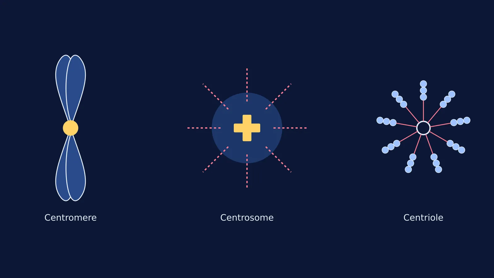
When a cell prepares to divide, its DNA condenses into visible chromosomes. A replicated chromosome looks like an 'X', made of two identical halves called sister chromatids. The centromere is the pinched, constricted region of DNA where these two chromatids are held together.
According to the National Council of Educational Research and Training (NCERT), the centromere is known as the "primary constriction" of the chromosome. You will find disc-shaped protein structures called kinetochores sitting on the sides of the centromere. During cell division, spindle fibres attach to these kinetochores—not to the DNA itself—to pull the chromatids apart. Chromosomes are classified into four types (metacentric, sub-metacentric, acrocentric and telocentric) based entirely on where the centromere is positioned. If you need to revise these shapes, you can review the chromosome types by centromere position.
What is a centrosome?
The centrosome is an organelle located in the cytoplasm, far away from the DNA. It acts as the main microtubule organising centre (MTOC) for animal cells.
While higher plant cells manage to divide without them, almost all animal cells rely on centrosomes to build the spindle apparatus. During the S-phase of the cell cycle, the centrosome duplicates in the cytoplasm while the DNA is replicating inside the nucleus. When mitosis begins in prophase, the two centrosomes move to opposite poles of the cell and cast out a net of microtubule fibres. These fibres reach out, grab the chromosomes by their kinetochores, and pull them into the two new daughter cells.
If you find yourself memorising these steps with concept gaps, the Fermi Concept Library covers every concept in biology, each with its own lesson to help you map out where organelles move during cell division.
What is a centriole?
If you zoom in on a centrosome, you will find that it is not a solid sphere. It is made of a cloudy protein matrix surrounding two tiny, barrel-shaped structures. These cylinders are the centrioles.
In a centrosome, the two centrioles lie perpendicular to each other. They are built entirely out of a protein called tubulin. When viewed in cross-section, a centriole looks like a cartwheel. It has a central protein hub connected by radial spokes to nine peripheral fibrils. Each fibril is made of three microtubules grouped together. This is famously known as the "9+0" arrangement, because there are nine triplets on the outside and zero microtubules in the dead centre (NCERT).
Beyond helping cells divide, centrioles have a second vital job: they migrate to the cell membrane to form basal bodies. These basal bodies act as the roots for cilia and flagella and the 9 + 2 axoneme structures that help cells move or sweep fluid across their surface.
Centromere vs centrosome vs centriole: A cheat sheet for NEET
The easiest way to lose marks on a biology paper is to read one of these words and mistake it for another. Here is how to keep them strictly separated in your mind:
Centromere: The DNA region on a chromosome. It holds chromatids together and anchors the kinetochore. Every eukaryotic cell has them.
Centrosome: The organelle in the cytoplasm. It organises the spindle fibres. It is found in animal cells, but missing in higher plants.
Centriole: The protein cylinder. There are two inside every centrosome. They form the 9+0 cartwheel structure and create basal bodies.
The "9+0" vs "9+2" trap
A classic student mistake is mixing up the microtubule arrangements of centrioles and cilia.
Because centrioles give rise to the basal bodies of cilia, it is tempting to assume they share the exact same internal architecture. They do not. Centrioles have a 9+0 arrangement (nine triplets on the edge, an empty protein hub in the middle). Cilia and flagella, however, have a 9+2 arrangement (nine doublets on the edge, plus two distinct microtubules running down the centre). When an exam question asks about the "cartwheel" model, it is always referring to the 9+0 centriole, never a cilium. Keeping this detail straight will save you from one of the most common traps in multiple-choice exams.


