

On this page
Once a cell has successfully duplicated and separated its chromosomes (karyokinesis), it is still a single entity. To finish the process and become two independent daughter cells, it must divide its cytoplasm and all the organelles floating within it. This final act of the cell cycle, mitosis and meiosis is called cytokinesis.
While the goal—creating two separate cells—is identical across most forms of life, the mechanism differs sharply between plants and animals. This difference comes down to one defining architectural feature: the rigid plant cell wall.
How does cytokinesis happen in animal cells?
Animal cells are bound only by a flexible, fluid plasma membrane. Because they lack a rigid outer shell, they can achieve cytokinesis by simply pinching themselves in two.
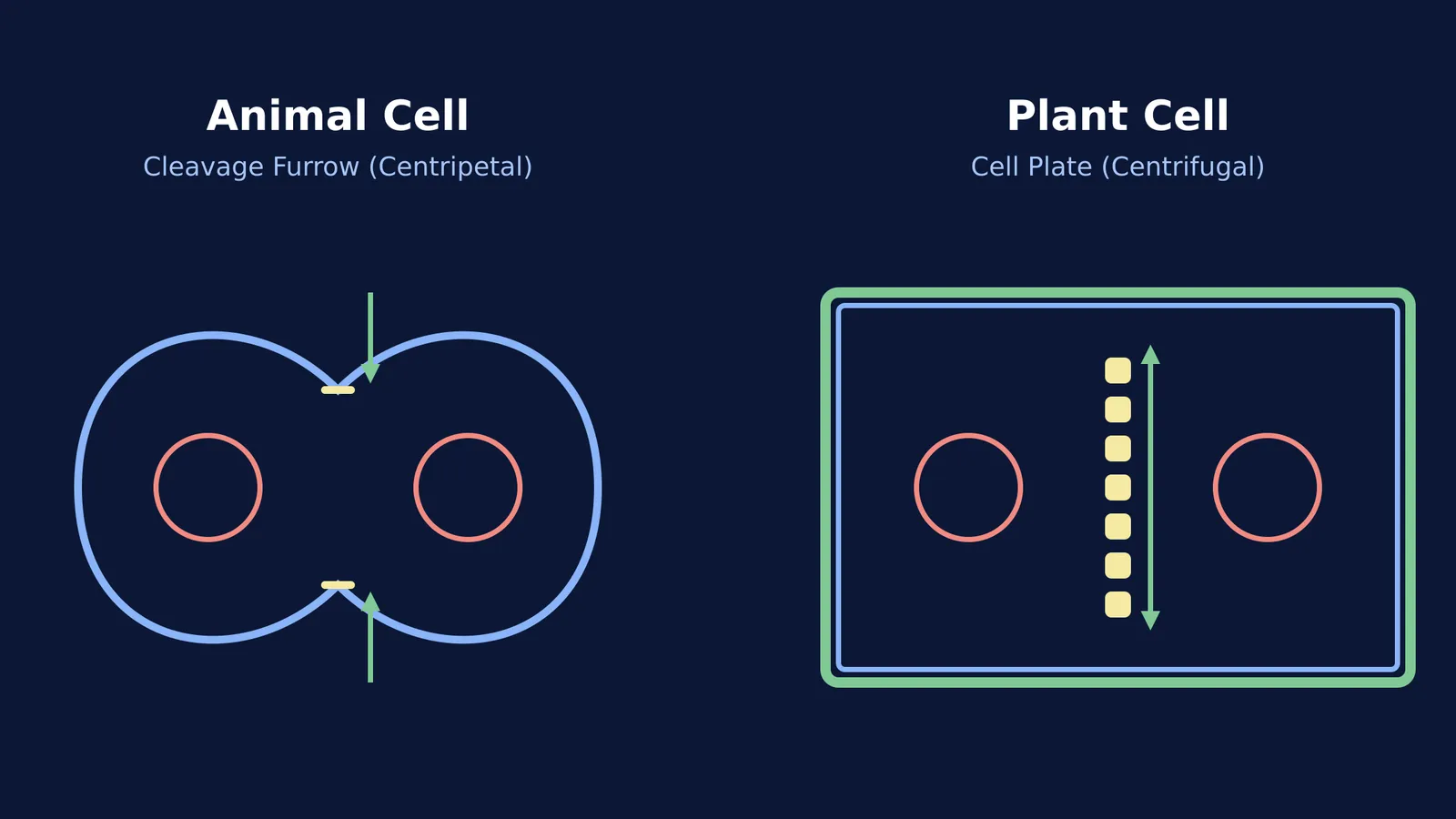
The cleavage furrow: Cytokinesis begins when a shallow groove, known as a cleavage furrow, appears in the plasma membrane at the cell's equator.
The contractile ring: Just beneath the membrane lies a ring of microfilaments made of actin and myosin. These are the same proteins responsible for muscle contraction. As these filaments slide past one another, the ring tightens, pulling the cell membrane inward like the drawstring of a bag.
Centripetal division: Because the furrow starts at the outer edge (the periphery) and moves inward toward the center, this type of division is described as centripetal.
The furrow gradually deepens until the membrane meets in the middle and fuses. The parent cell's cytoplasm is completely split, leaving two distinct daughter cells.
What is the difference in plant cell cytokinesis?
Plant cells are enclosed by a tough, inextensible cell wall made largely of cellulose. Because of this rigid armor, the cell cannot simply pinch inward without shattering. Instead, it must build a new dividing wall from the inside out.
The cell plate: Rather than a furrow, plant cells form a precursor structure called the cell plate. Vesicles dispatched from the Golgi apparatus gather at the center of the cell, carrying materials needed to build a cell wall.
The middle lamella: These vesicles fuse together at the equator. The resulting cell plate represents the middle lamella—the sticky layer (made mostly of calcium pectate) that will glue the walls of the two adjacent plant cells together.
Centrifugal division: The cell plate starts at the center and grows outward toward the lateral walls until it fuses with the existing plasma membrane. Because the division spreads from the inside to the outside, it is called centrifugal.
As this central wall forms, organelles such as mitochondria and plastids are distributed roughly equally between the two newly forming daughter cells.
If you struggle to picture how these structures assemble, you can map out the pathways in the Fermi Concept Library, or ask Fermi's AI tutor to draw the figures and explain the process step by step.
What happens when karyokinesis is not followed by cytokinesis?
In standard cell division, cytokinesis happens almost immediately after the nucleus divides. However, biology is full of exceptions. In some organisms and specific tissues, karyokinesis (nuclear division) occurs repeatedly without ever triggering cytokinesis (cytoplasmic division).
When the nucleus divides but the cell does not split, the result is a single, large cell containing multiple nuclei. This multinucleate condition is called a syncytium.
The classic NCERT example of a syncytium is the liquid endosperm of a coconut—the sweet coconut water inside. After fertilisation, the primary endosperm nucleus undergoes rapid, successive free-nuclear divisions without forming cell walls between the new nuclei. This creates a massive liquid syncytium containing thousands of free-floating nuclei. Over time, cell walls begin to form from the periphery inward, creating the white, fleshy cellular endosperm (the coconut meat), but the liquid center remains a syncytium.
Biologists have studied these decoupled divisions since the days of Walther Flemming, noting that skipping cytokinesis is often an evolutionary strategy. It allows an organism to replicate DNA and grow a tissue mass rapidly without spending the immense energy required to build thousands of individual cell membranes and walls.
How do you compare plant and animal cytokinesis in exams?
When revising this topic for biology exams, make sure you can answer these four comparative points clearly:
Direction of division: Is it centripetal (animal) or centrifugal (plant)?
Primary structure formed: Does the cell build a cleavage furrow (animal) or a cell plate (plant)?
Role of the cell wall: Mention that the flexible membrane allows furrowing in animals, while the rigid wall forces plate formation in plants.
Organelle involvement: Note that animal cytokinesis relies on actin-myosin microfilaments, whereas plant cytokinesis relies heavily on Golgi vesicles transporting pectin.
Mastering this distinction ensures you are ready for both standard comparison questions and deeper questions on multinucleate cell anomalies.


