
On this page
Prophase, metaphase, anaphase, telophase: every biology student learns the stages of mitosis. The word mitosis itself, and the word chromatin, come from one book written in 1882 by a German anatomist who spent years watching cells divide. This post covers who Walther Flemming was, what he saw and named, a short calculation about how often you catch a cell dividing, common mistakes, and what to learn from his methods.
Who was Walther Flemming?
The Deutsche Biographie records that Flemming was born on 21 April 1843 at Sachsenberg near Schwerin and died in Kiel on 4 August 1905. He studied medicine at Göttingen, Tübingen, Berlin and Rostock, served as a doctor in the war of 1870-71, worked in Prague, and in 1876 became professor of anatomy at Kiel, retiring through illness in 1901.
How did he study dividing cells?
The same entry explains his method. He combined observations of living material, tissue from salamander larvae, with fixed and stained preparations. Watching living cells showed the order of events; staining showed the detail. He also improved fixing and staining, and introduced a fixative of chromic, osmic and acetic acids still known as Flemming's solution.
He saw that before division the stainable material in the nucleus forms characteristic threads (the NDB calls them loops), which Wilhelm Waldeyer later named chromosomes. He described how each thread splits lengthwise and how the halves form two new nuclei. Following the botanist Eduard Strasburger, he stressed that cell division is basically the same in plants and animals.
Where do "chromatin" and "mitosis" come from?
Both appear in his 1882 book Zellsubstanz, Kern und Zelltheilung (Cell substance, nucleus and cell division).
On chromatin he wrote, in our translation, that he used the name "quite empirically" for "the substance in the cell nucleus that takes up the colour when nuclei are stained". He kept the name, he said, until chemistry could tell what the substance really was.
In a chapter on names he proposed replacing "karyokinesis", which only means movement in the nucleus, with Karyomitosis, which "briefly expresses: thread metamorphosis in the nucleus". He suggested calling the division figures simply "Mitosen" and division without thread changes "amitotic". The Greek mitos means thread.
He was modest about it. He called new terms "a necessary evil" and wrote that "the essential thing is the observations, not the words with which one describes them." He said he would not use his new names himself until others took them up.
Worked example: how often do you catch a cell in mitosis?
NCERT's Cell Cycle and Cell Division chapter says human cells in culture divide about once every 24 hours, and division proper lasts only about an hour. Interphase takes more than 95 per cent of the cycle.
Fraction in M phase. If cells are at random points in the cycle, the fraction dividing at any moment is about 1 h ÷ 24 h = 0.042, or 4.2 per cent.
Interphase share. 23 ÷ 24 = 95.8 per cent, which matches NCERT's "more than 95%".
In a field of cells (sample number). Look at 1,200 such cells and you would expect about 1,200 × 1/24 = 50 in some stage of mitosis.
This is why Flemming needed so many preparations, and why fast-growing tissue such as an onion root tip is used in the school practical: more cells are dividing. Our post on the onion root tip practical covers the method.
What mistakes do students make with mitosis?
Confusing chromatin and chromosomes. Chromatin is the stainable material; chromosomes are what it condenses into during division.
Counting DNA and chromosomes the same way. After S phase, DNA goes from 2C to 4C but chromosome number stays 2n, as NCERT notes.
Calling interphase "resting". NCERT points out the cell is preparing for division the whole time.
Treating 4.2 per cent as universal. It assumes a 24-hour cycle and cells spread evenly through it.
For the full stage-by-stage sequence, see our post on the cell cycle, mitosis and meiosis. If a moving process is hard to follow from still diagrams, the Fermi Simulation Lab has interactive simulations for ideas easier to see than read.
What can you take from how Flemming worked?
Use two views of the same thing. Living cells gave order; stained cells gave detail.
Improve your tools. His fixative and staining work made the observations possible.
Define words by what you can test. His chromatin was "what takes the dye", nothing more.
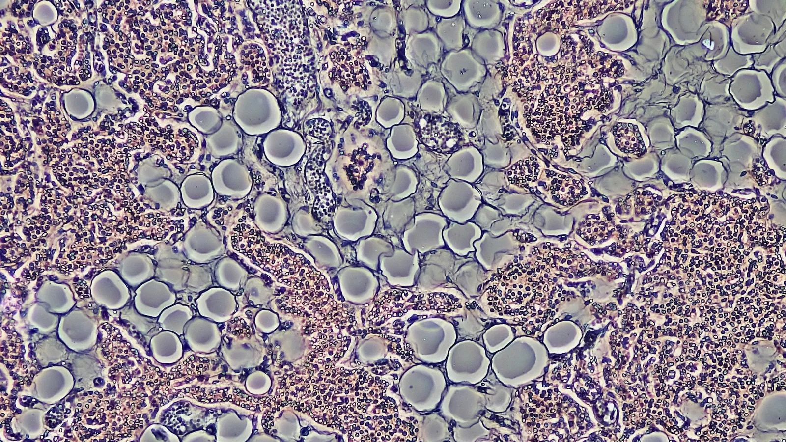
Next step: count dividing and non-dividing cells in a textbook micrograph of a root tip and calculate the fraction yourself.