

On this page
The onion root tip slide is one of the experiments you actually make, not just look at. It is also one where students often see a pink smear and no chromosomes. This post explains why each step of the method exists, how to recognise each stage, and how to turn your cell counts into a mitotic index for your record and viva.
Where is this in the CBSE practical exam?
In the CBSE Biology curriculum for 2026-27, the Class 12 list of experiments includes "Prepare a temporary mount of onion root tip to study mitosis". Slide preparation, marked out of 5, covers this experiment and the pollen germination slide. The 3-hour practical exam is worth 30 marks in total. In Class 11, mitosis in onion root tip cells appears as a spotting item from permanent slides, so you may have seen the stages already. Check your school's schedule for the current year.
Why use onion root tips?
Mitosis happens where cells are being made. NCERT's cell cycle chapter says mitotic divisions in plants take place in meristematic tissues, such as the apical meristems, and a root tip has an apical meristem just behind its cap. Onion bulbs grow fresh roots quickly in water, and the same chapter notes that onion root tip cells have 16 chromosomes, few enough to see.
Onion is also a research tool. In a 1985 paper in Hereditas, Geirid Fiskesjö proposed the "Allium test" as a standard in environmental monitoring, citing its low cost, ease of handling and good chromosome conditions for studying chromosome damage and disturbed cell division. Your practical uses the same material for a simpler job.
What does each step of the method do?
Your teacher or lab manual will give exact chemicals, strengths and timings. Know the purpose of each step, because viva questions ask "why", not "how long".
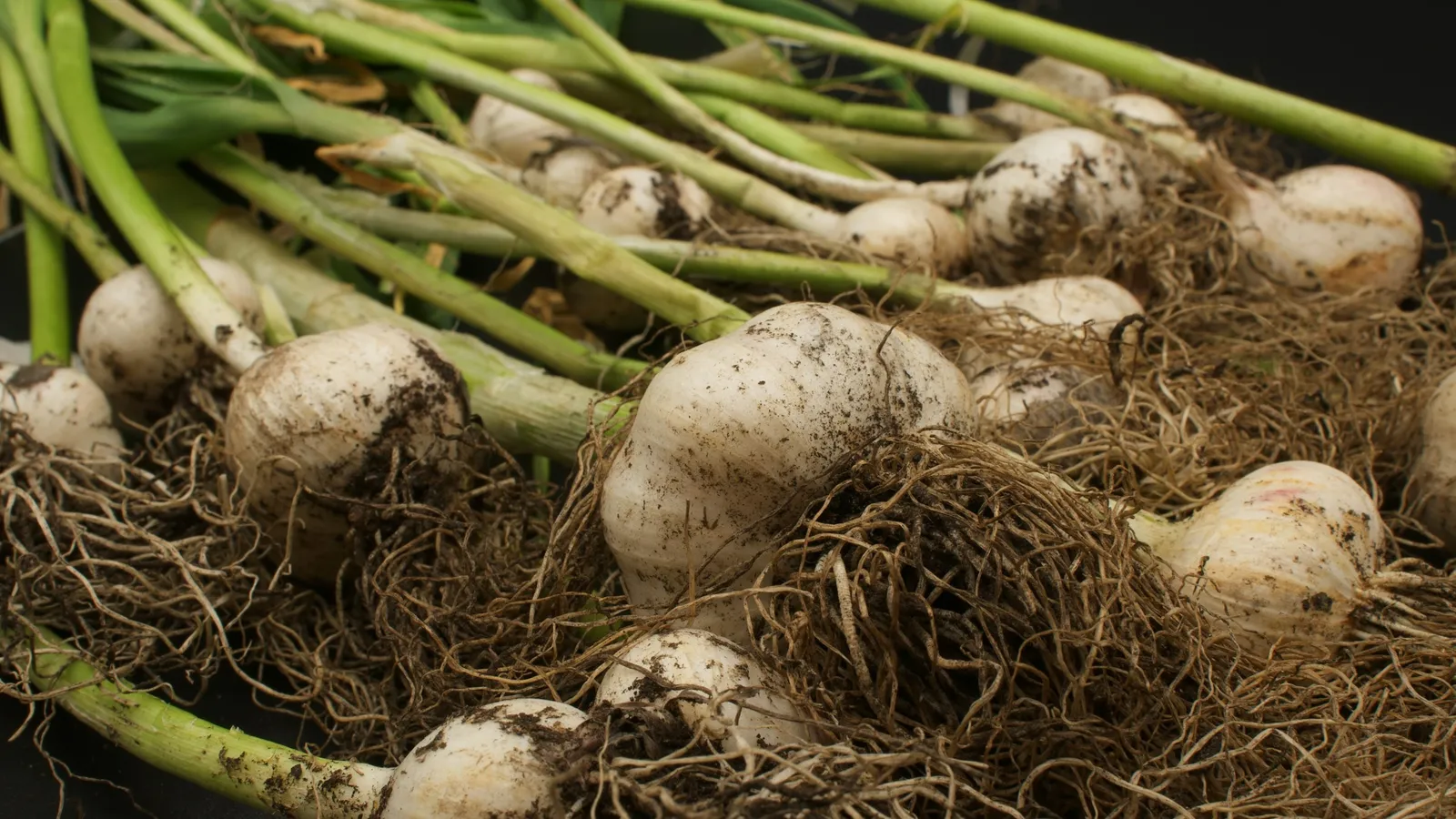
Grow roots. Rest an onion bulb on a jar of water so its base just touches. New roots appear within a few days.
Cut the tips. Take only the last few millimetres, where the meristem is.
Fix. A fixative stops the cells where they are, so dividing cells stay at their stage.
Soften with acid. Warm dilute hydrochloric acid breaks down the material that holds cells together, so they can be spread into a single layer.
Stain. A chromosome stain such as acetocarmine colours the chromosomes darker than the cytoplasm.
Squash. Place a coverslip and press gently and evenly, without sliding it. You want a single layer of cells, not a torn mess.
Observe. Find the cells under low power, then switch to high power for the stages.
How do you identify the stages?
Use the features NCERT gives:
Interphase: an intact nucleus with a nucleolus; no visible chromosomes. Most cells look like this.
Prophase: chromosomes condense into visible threads; the nuclear envelope is still present early on.
Metaphase: condensed chromosomes line up at the equator, on the metaphase plate. NCERT says this is when chromosome shape is easiest to study.
Anaphase: sister chromatids separate and move to opposite poles.
Telophase: chromosomes cluster at the poles and a nuclear envelope forms around each group. In plant cells, NCERT says the new wall starts as a cell plate in the centre and grows outward.
Worked example: mitotic index from sample counts
The numbers below are sample numbers for practice, not results from a real slide. A student counts 400 cells in several fields of view:
Interphase: 328
Prophase: 48
Metaphase: 10
Anaphase: 6
Telophase: 8
Dividing cells = 48 + 10 + 6 + 8 = 72. Check: 328 + 72 = 400.
Mitotic index = dividing cells / total cells × 100 = 72 / 400 × 100 = 18%.
The share of each stage among dividing cells: prophase 48 / 72 ≈ 67%, metaphase 10 / 72 ≈ 14%, anaphase 6 / 72 ≈ 8%, telophase 8 / 72 ≈ 11%. A stage that appears more often generally lasts longer, so in these counts prophase is the longest stage and anaphase the shortest.
Why is the index well below half? NCERT notes that in a typical human cell, interphase lasts more than 95% of the cell cycle. Expect most root tip cells to be in interphase too, even in a busy meristem.
Which mistakes cost marks?
Over-pressing the coverslip and smearing the cells.
Calling a densely stained interphase nucleus prophase. Look for separate threads.
Drawing what the textbook shows instead of what your slide shows. Draw one real cell per stage.
Writing "mitosis seen" in your record without counts. A short table and a mitotic index show you observed.
For the theory behind the stages, read our post on cell cycle, mitosis and meiosis, and for the viva, board practical exams and viva questions. If the order of events in mitosis still feels like a list to memorise, Fermi's AI tutor draws the figure and checks your understanding as it goes, so you can test yourself stage by stage.
A practice routine before the exam
Draw the five stages from memory and label one feature each.
Explain each step of the method in one sentence, starting with "so that".
Work out a mitotic index from your own slide or the sample counts above.
Ask a friend three "why" viva questions: why the tip, why acid, why gentle pressure.
Before your practical, set an onion on a jar of water so you see its roots grow for yourself.