On this page
"Identify the stage shown" is a familiar question in Class 11 biology and NEET-UG practice. Many students know each stage in words but mix up the pictures, especially the three metaphases. Instead of matching against one memorised picture, read three clues every diagram gives you, in a fixed order.
Everything below follows chapter 10, "Cell Cycle and Cell Division", of the NCERT Class 11 Biology textbook, with the descriptions cross-checked against OpenStax Biology 2e.
What clues tell you the stage of cell division?
Look for these three things, in this order:
Nuclear envelope. In mitosis the envelope starts to break into small vesicles in prophase and forms again in telophase, according to OpenStax Biology 2e. Thin threads inside an intact nucleus mean early prophase. New envelopes forming around two clusters at the poles means telophase.
Chromosome position. Chromosomes scattered through the cell mean prophase. A line across the middle means metaphase. Chromosomes on their way to the poles mean anaphase. Chromosomes bunched at the poles mean telophase.
Pairing and chromatids. Homologous chromosomes lying side by side mean meiosis I. Single X-shaped chromosomes, each made of two sister chromatids, mean mitosis or meiosis II.
The NCERT chapter adds an anaphase clue: each centromere leads, "with the arms of the chromosome trailing behind", so its Figure 10.2 draws them as V shapes.
How do you tell mitotic metaphase from metaphase I and metaphase II?
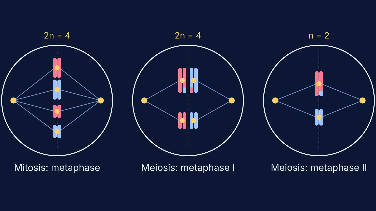
All three show chromosomes at the middle of the cell, so position alone won't settle it. Look at how they are lined up.
Mitotic metaphase: the chromosomes form one single-file line across the middle. Each one has two sister chromatids, and spindle fibres from opposite poles attach to the two kinetochores of the same chromosome (NCERT, section 10.2.2). In a diploid cell you will see two of each size and shape, but they are not paired.
Metaphase I: the line is double. NCERT says that "the bivalent chromosomes align on the equatorial plate". Each bivalent is a pair of homologous chromosomes, four chromatids in all. OpenStax adds that the homologs' kinetochores face opposite poles. In a diagram, each partner of a pair has its own spindle fibre coming from one side.
Metaphase II: the line is single again, as in mitosis, but the cell holds only one set of chromosomes. OpenStax puts it this way: meiosis II works like mitosis "except that each dividing cell has only one set of homologous chromosomes, each with two chromatids". Each size and shape appears once. NCERT's Figure 10.4 draws two such cells, one above the other, because meiosis I has already made two cells.
The same logic works for anaphase. In anaphase I the homologs separate, and NCERT notes that sister chromatids "remain associated at their centromeres", so whole X-shaped chromosomes move to each pole. In mitotic anaphase and anaphase II, the centromere splits and single chromatids move.
How do you spot the stages of prophase I?
NCERT divides prophase I into five substages: leptotene, zygotene, pachytene, diplotene and diakinesis (NCERT, section 10.4.1). Use these diagram clues:
Thin, single threads that are only just visible: leptotene.
Homologs starting to pair up (synapsis): zygotene.
Thick bivalents with four clear chromatids: pachytene, when crossing over takes place.
Homologs pulling apart but still joined at X-shaped chiasmata: diplotene.
Chiasmata moving to the chromosome ends while the nucleolus and nuclear envelope disappear: diakinesis.
A worked example: reading one diagram
Say a question shows a cell with no nuclear envelope. Four X-shaped chromosomes sit in a double row across the middle, and spindle fibres from the left pole reach only the left-hand row.
Envelope gone, chromosomes in the middle: some kind of metaphase.
Double row, with partners of the same size and shape facing each other: homologs are paired, so these are bivalents.
Each pole connects to just one homolog of each pair: this matches metaphase I.
Now change one detail: the four chromosomes are in single file, and there are two long and two short ones. That's mitotic metaphase. If there's one long and one short chromosome in single file, it's metaphase II. For the counts behind each stage, see our guide to chromosome number and DNA content through mitosis.
How should you practise for exams?
Assertion-reason questions test the same distinctions, as our post on cell division assertion-reason traps shows. Our explainer on how meiosis halves chromosomes and creates variation explains why pairing in meiosis I matters at all.
If a term like bivalent, kinetochore or chiasma is still unclear, the Fermi Concept Library has a lesson on every concept in biology and shows where it fits in the map. For practice, the Fermi Problem Library has worked problems with a step-by-step solver that explains the reasoning rather than printing an answer.