
On this page
If you are revising the cell chapter for CBSE or NEET-UG, microvilli and cilia are easy to mix up. Both stick out from the free surface of epithelial cells, and both look like a fringe in a textbook diagram. The quickest test: does it move? Cilia beat; microvilli do not.
What are microvilli and what do they do?
A brush border is an array of actin-supported membrane protrusions called microvilli that increases the functional capacity of the tissue, according to a 2014 review in the Journal of Cell Biology by Crawley, Mooseker and Tyska.
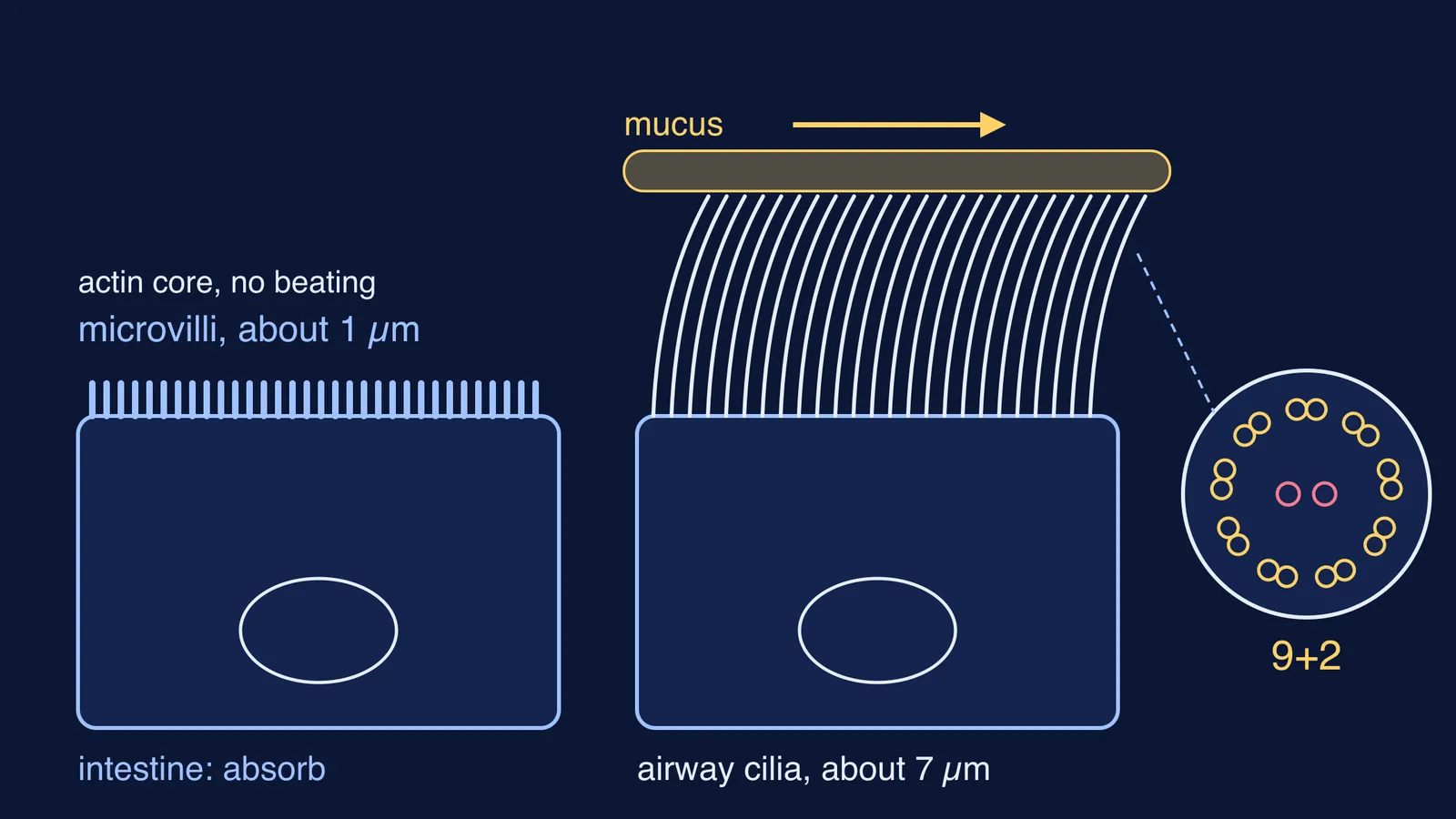
Microvilli are about 1 µm long, and the small intestine carries an estimated 200 million of them per square millimetre, says the OpenStax Anatomy and Physiology chapter on the intestines. Together they look like a mass of bristles, hence the name brush border. Enzymes fixed to the microvillus membrane finish digesting carbohydrates and proteins right where the food is absorbed.
The same chapter says circular folds, villi and microvilli together increase the absorptive surface more than 600-fold. For what happens to fats once they cross this surface, see our explainer on how fats are absorbed through micelles, chylomicrons and lacteals.
What are cilia and what is the 9+2 axoneme?
NCERT's Class 11 chapter Cell: The Unit of Life describes cilia as hair-like outgrowths of the cell membrane that work like oars, moving either the cell or the fluid around it. Their core, the axoneme, usually has nine doublets of peripheral microtubules around a pair of central microtubules: the 9+2 array. Cilia emerge from a centriole-like basal body.
A review by Satir and Christensen in the Annual Review of Physiology describes the motile kind: in the multiciliated cells of mammalian epithelia, axonemes are 9+2, carry dynein arms and are motile. The same review describes single, non-motile 9+0 primary cilia on cells such as those of the kidney tubule, so not every cilium beats.
Mammalian airway cilia are around 7 µm long, packed 100 to 200 per cell, and beat at 7 to 20 Hz, according to Sears and colleagues in the American Journal of Physiology. Set beside the 1 µm microvillus from OpenStax, a typical airway cilium is roughly seven times longer. Our guide to micrometres and nanometres for cells and membranes puts them on one scale.
Where are microvilli and cilia found in the body?
Microvilli: on the absorptive cells lining the small intestine, where they form the brush border (OpenStax, small and large intestines).
Cilia in the airway: ciliated epithelium forms a mucociliary escalator that sweeps dust and pathogens trapped in mucus toward the throat (OpenStax, epithelial tissue).
Cilia elsewhere: the lining of the fallopian tubes, and the ventricles of the brain, where they help circulate cerebrospinal fluid (same OpenStax section).
Much of the airway is lined by pseudostratified columnar epithelium, in which some cells carry cilia. Single-celled organisms use cilia too; see Paramecium's cilia and oral groove.
Microvilli vs cilia: what is the difference, point by point?
Core: microvilli are supported by actin; cilia have a microtubule axoneme in a 9+2 pattern, rising from a basal body.
Size: microvilli about 1 µm (OpenStax); mammalian airway cilia about 7 µm (Sears and colleagues).
Movement: microvilli do not beat; motile cilia beat, and their axonemes carry dynein arms.
Job: microvilli add surface for absorption and carry digestive enzymes; cilia move mucus, fluid or the whole cell.
Where: microvilli on the intestinal lining; cilia in the airways, fallopian tubes and brain ventricles.
How do you draw and label microvilli and cilia in an exam?
Worked example: draw a labelled diagram comparing the two in five steps, as in the sketch at the top (lengths from OpenStax and Sears and colleagues):
Draw two tall columnar cells side by side, each with a nucleus near the base.
On the left cell, draw a dense row of short, even projections. Label it microvilli (brush border) and write absorption under it.
On the right cell, draw fewer, longer, curved projections, roughly seven times the microvillus height. Label cilia and basal body.
Draw a mucus layer resting on the cilia tips, with an arrow showing which way it is swept.
Beside the cilia, add a small circle with nine pairs around two central dots, labelled 9+2 axoneme.
Then check your answer against three common traps:
Calling microvilli tiny cilia. Their core is actin, not an axoneme, and they do not beat.
Writing 9+2 for every cilium. Primary cilia are 9+0 and non-motile.
Mixing up villi and microvilli. Microvilli are much smaller than villi and project from one cell's plasma membrane.
If a definition still feels shaky, the Fermi Concept Library gives every biology concept its own lesson and its place in the map. For practice, the Fermi Problem Library has worked problems with a step-by-step solver that explains the reasoning rather than printing an answer.
Tonight, draw the sketch from memory and check it against the five steps.