On this page
Paramecium is one of the protists you meet in Class 11 Biology, and it keeps turning up in questions on Kingdom Protista. Here is the cell from the outside in, then a plan for drawing it without losing marks.
What does Paramecium look like under a microscope?
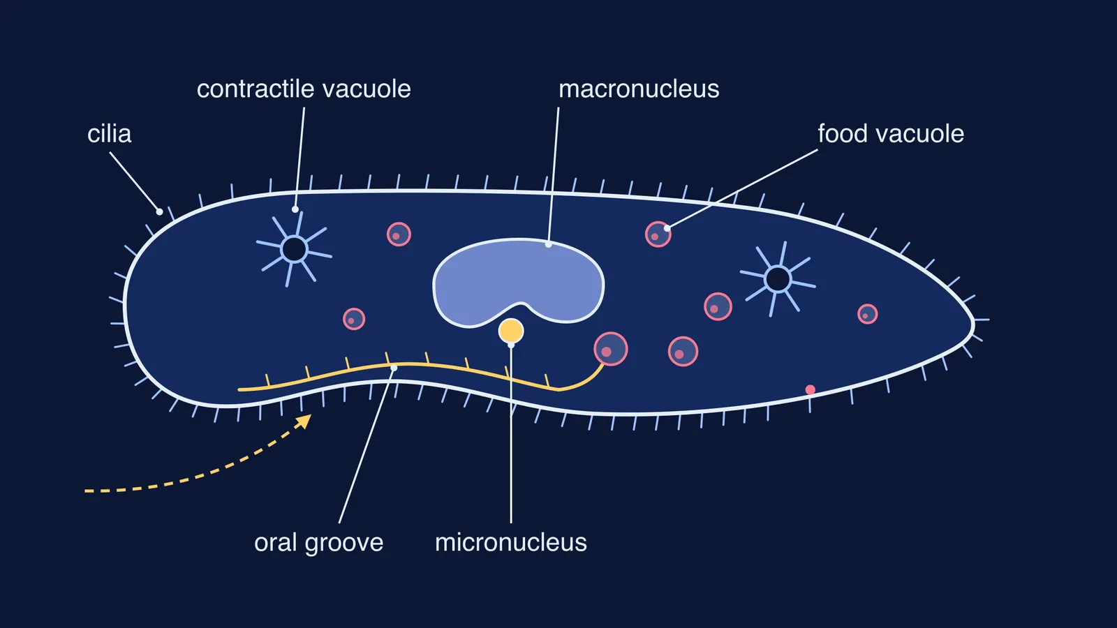
Paramecium caudatum, a common species, is typically 170 to 330 micrometres long, with a rounded front end and a body that tapers to a blunt point at the back (Freshwater Biodiversity Portal of Bangladesh). That outline, plus the dent of the oral groove along one side, is why it is called slipper-shaped.
NCERT's Class 11 Biology textbook groups Paramecium with the ciliated protozoans: aquatic organisms that move actively "because of the presence of thousands of cilia" (NCERT, Biological Classification).
How do cilia help Paramecium move and feed?
Cilia are short, hair-like projections that beat like tiny oars. In Paramecium they row the cell through water and also set up currents that carry food towards it.
NCERT puts the feeding job in one line: coordinated movement of rows of cilia steers water laden with food into the gullet, a cavity that opens to the outside of the cell (NCERT, Biological Classification).
High-speed video of swimming Paramecium found that most cilia beat 15 to 45 times a second, while cilia in the region around the mouth beat at almost double that rate (Funfak et al., Integrative Biology, 2015). Faster beating went with faster swimming, in a nearly straight-line relationship.
What is the oral groove in Paramecium?
The oral groove is a long depression on one side of the cell that leads into a deeper oral cavity lined with cilia (Freshwater Biodiversity Portal of Bangladesh). Think of it as a funnel: cilia sweep food particles down the groove, through the gullet, to the cell mouth (cytostome).
At the bottom of the gullet, a new food vacuole pinches off, carrying its load of food into the cytoplasm (Richard Allen's Paramecium atlas, University of Hawaii). In Paramecium caudatum, the pH inside new food vacuoles fell from about 7 to 3 within 5 minutes of their formation (Fok, Lee and Allen, Journal of Protozoology, 1982; abstract on the NIES protozoa database). No labelled vacuole was emptied before 21 minutes, and almost all were emptied within an hour, so each digestive cycle took 21 to 60 minutes.
Leftovers leave through a fixed spot on the surface, the anal pore or cytoproct, and contractile vacuoles pump out water that enters by osmosis (Wikipedia, Paramecium).
The two contractile vacuoles and their radiating canals are described on the Freshwater Biodiversity Portal; our explainer on how the contractile vacuole works in Amoeba and Paramecium covers how they fill and empty.
Why does Paramecium have a macronucleus and a micronucleus?
Paramecium splits the work of a nucleus in two. A review of Paramecium genetics describes two diploid micronuclei that undergo meiosis and fertilisation to pass genetic information to the next sexual generation, and a highly polyploid macronucleus holding a trimmed genome set up for gene expression (Duharcourt and Sperling, Methods in Enzymology, 2018). The number of micronuclei varies: a cell has one or more, depending on the species (Wikipedia, Paramecium).
As the macronucleus develops, its DNA is copied until it holds about 800 copies of the genome, according to the same review. Put simply: the macronucleus runs the cell day to day; the micronucleus is the copy kept for reproduction.
For contrast, Amoeba has no cilia and catches food with false feet; see how Amoeba feeds using pseudopodia.
How do you draw and label Paramecium for exams?
Draw in this order and you will not miss a part:
Outline: rounded front, tapering back, much longer than it is wide.
Oral groove cut into one side, from near the front to about the middle.
A short gullet at the end of the groove, with a food vacuole forming at its tip.
A large, bean-shaped macronucleus near the centre and a small micronucleus beside it.
Two star-shaped contractile vacuoles, one towards each end.
A few food vacuoles of different sizes in the cytoplasm.
Short cilia all round the edge, then labels with straight, parallel lines.
Before you hand it in, check:
Is the oral groove on the side, not at the tip?
Did you draw two contractile vacuoles?
Did you write the function beside macronucleus and micronucleus, not just the name?
Are the cilia short and even, not long whips like a flagellum?
Our guide to making a hay infusion and watching protozoa multiply shows how to set one up.
If terms like cilia, osmoregulation and polyploidy still blur together, the Fermi Concept Library gives each concept its own lesson and its place in the map. To practise the diagram with feedback, Fermi's AI tutor can draw the figure with you and check your understanding as it goes.