
On this page
Fat is the awkward nutrient. Sugars and amino acids dissolve in water, but fat does not. So the body takes fat apart, ferries the pieces across, rebuilds it inside the villus cells, wraps it in protein, and sends it along a side road: the lymph.
How are fats digested before they can be absorbed?
Most dietary fat is triglyceride: one glycerol with three fatty acids attached. In the small intestine, bile and pancreatic juice supply pancreatic lipase, bile salts and colipase, which work together to digest and absorb lipids, according to a review by Iqbal and Hussain in the American Journal of Physiology (2009). Bile salts help break fat into smaller droplets (emulsification), giving lipase more surface; our post on how bile emulsifies fat covers that step.
Pancreatic lipase cuts the fatty acids at positions 1 and 3 of each triglyceride, releasing 2-monoglyceride and free fatty acids, the same review explains. These products gather with cholesterol, phospholipids and bile salts into mixed micelles: tiny clusters with a water-facing surface and a fatty core. The review notes that bile salt micelles help cholesterol cross the still layer of water next to the cell surface.
What happens to fat inside the villus cells?
The cells lining each villus are called enterocytes; our explainer on villi and absorption in the small intestine shows how they are arranged. Inside an enterocyte, most triglyceride is rebuilt along the monoglyceride pathway, in which monoglyceride and fatty acyl-CoA join to form diglyceride and then triglyceride, according to the Iqbal and Hussain review.
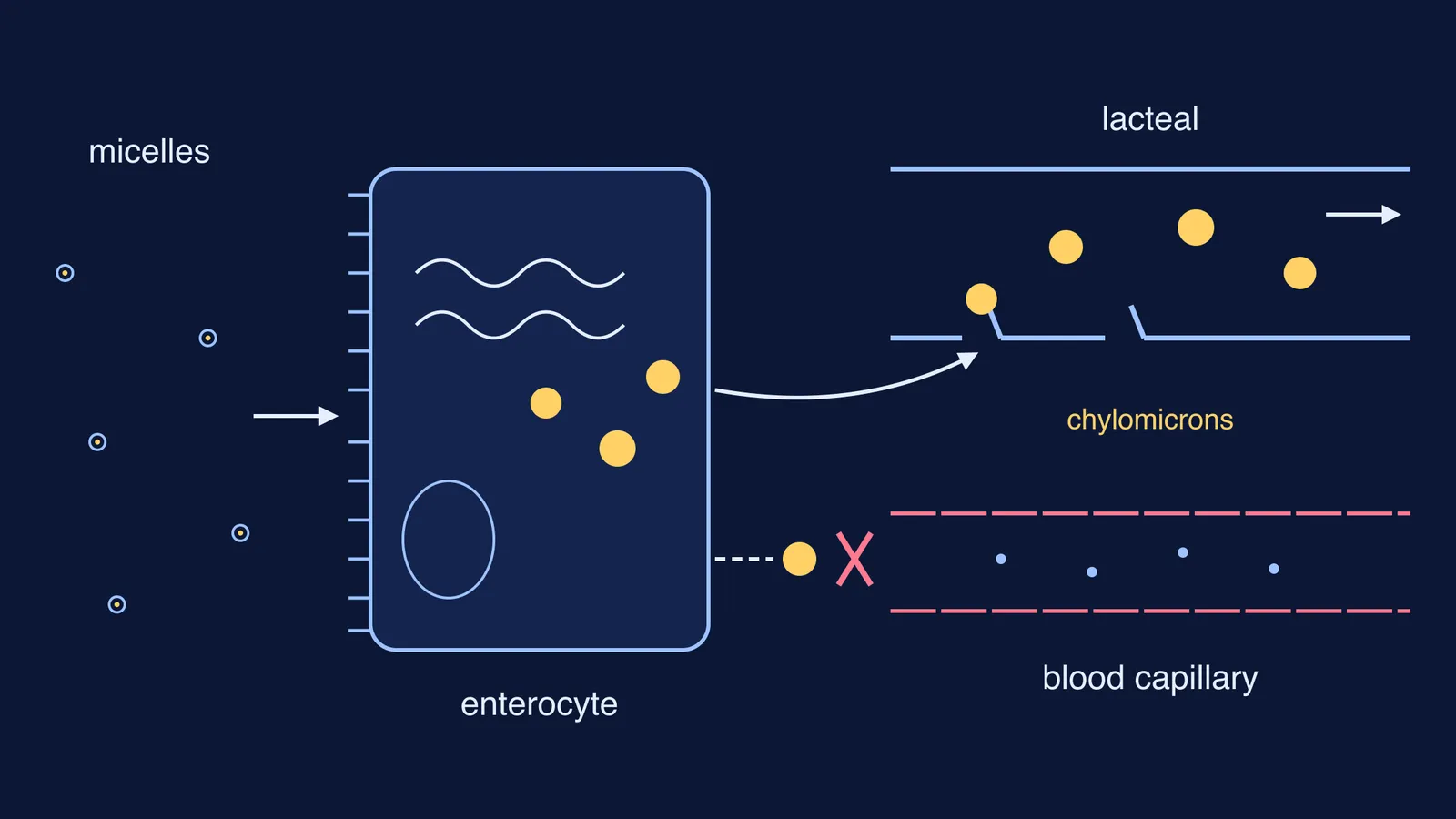
The new triglyceride is packed with cholesterol, phospholipid and a large protein called apoB48 into a chylomicron. The review calls apoB48 a key structural component of the chylomicron, and says chylomicrons are made only in the intestine, to carry dietary fat and fat-soluble vitamins. Finished chylomicrons leave from the base of the cell by exocytosis and move into the lamina propria, the tissue at the core of the villus, as described by Xiao and colleagues in Frontiers in Physiology (2020).
Why do chylomicrons enter lacteals instead of blood capillaries?
Each villus has a single central lymph vessel, the lacteal, surrounded by a network of blood capillaries. The short answer is size.
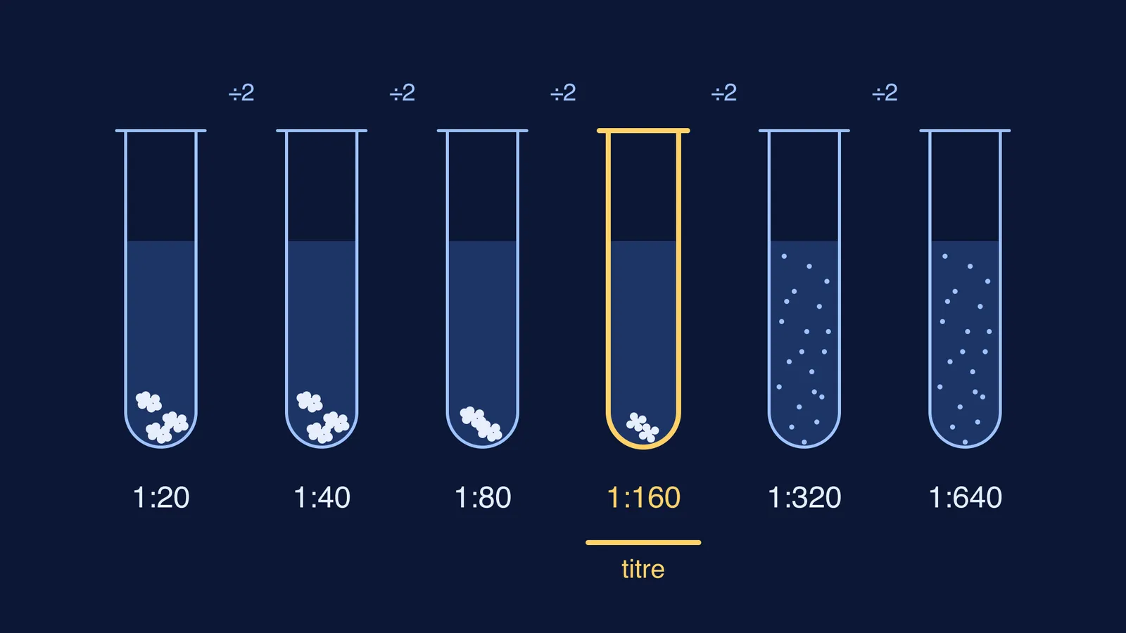
Chylomicrons are defined as particles at least 80 nanometres (800 angstrom) across, with an average size of about 160 nm, according to a review by Zhou, Qu, Liu and Tso in Frontiers in Physiology (2020).
The pores (fenestrations) in blood capillaries are “too narrow to permit passage of even the smaller size range of chylomicrons,” write Gwendalyn Randolph and Norman Miller in the Journal of Clinical Investigation (2014). The tip of the lacteal, by contrast, is thought to have pores large enough to let chylomicrons in, the same paper explains.
Xiao and colleagues add that most chylomicrons enter the lacteal and only a small fraction of very small ones may reach the blood capillaries. Lacteal walls have button-like junctions with gaps between them. In newborn mice, Zarkada and colleagues (Circulation Research, 2023) found that eaten lipids help open these junctions through a contraction signal called ROCK, while another signal, VEGF-A, works against the opening.
From the lacteals, chylomicrons travel through the intestinal lymph duct and the thoracic duct, and finally empty into the left subclavian vein, the Zhou review says. So fat packed in chylomicrons enters the blood in the upper chest rather than going straight to the liver through the hepatic portal vein, as most other nutrients do. For how lymph differs from blood plasma, see our post on lymph, plasma and tissue fluid.
One exception is worth knowing. Short- and medium-chain fatty acids are mostly carried unpackaged, bound to albumin, through the hepatic portal vein, while long-chain fatty acids are mostly packed into chylomicrons, the Zhou review notes.
How should you write this answer in an exam?
Use this five-step chain, each step with its reason:
Emulsify: bile salts break fat into small droplets, giving lipase more surface.
Digest: pancreatic lipase splits triglycerides into fatty acids and monoglycerides.
Ferry: micelles carry these pieces across the watery layer to the villus cells.
Rebuild and pack: enterocytes re-form triglycerides and coat them with protein, including apoB48, as chylomicrons.
Route: chylomicrons are too large for blood capillary pores, so they enter the lacteal and reach the blood through the thoracic duct.
Then check for these slips:
Writing that bile digests fat (it emulsifies; lipase digests).
Saying fatty acids enter the lacteal directly (they leave the cell inside chylomicrons).
Leaving out the thoracic duct.
Sending all absorbed fat to the liver through the portal vein.
If one step still feels shaky, the Fermi Concept Library has a lesson for every concept in biology, each with its place in the map. To practise the reasoning, the Fermi Problem Library has worked problems with a step-by-step solver that explains the reasoning rather than printing an answer.
Next step: draw one villus from memory, label the lacteal and the capillary, and write the five steps beside it.