

On this page
Every cell depends on extracellular fluid to deliver glucose, exchange gases, and remove metabolic waste. Yet biology questions often treat blood plasma, tissue fluid, and lymph as three unrelated liquids. In truth, they represent three consecutive stages of the same underlying fluid, separated by endothelial barriers and differing protein concentrations.
What is the difference between plasma, tissue fluid, and lymph?
The distinction between these fluids lies in where they reside and what they carry:
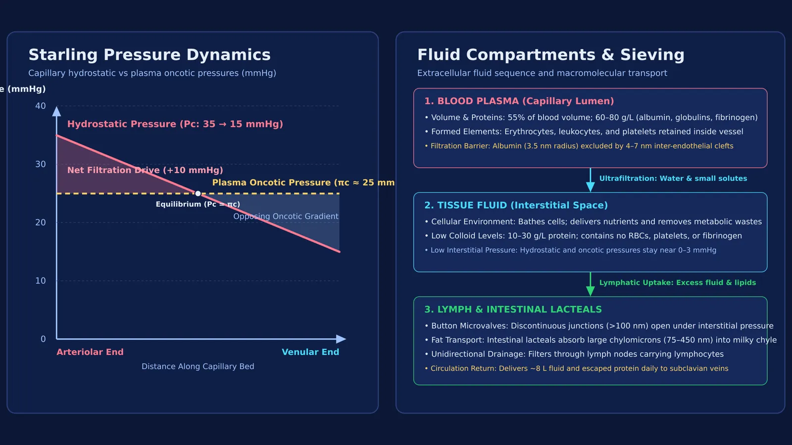
Blood plasma is the pale amber liquid matrix making up roughly 55 percent of total blood volume. As detailed in the StatPearls guide to blood plasma, it contains 6 to 8 percent dissolved proteins—mainly albumin, globulins, and fibrinogen—circulating alongside erythrocytes, leukocytes, and platelets described in our review of the formed elements of blood.
Tissue fluid, or interstitial fluid, bathes cells within microscopic tissue spaces. It forms when plasma filters across capillary walls, retaining water, dissolved gases, and small solutes while leaving cellular elements and large plasma proteins behind in the bloodstream.
Lymph is the clear fluid collected when excess interstitial fluid enters blind-ended lymphatic capillaries. According to StatPearls, lymph travels through lymph nodes carrying lymphocytes before returning to venous circulation near the heart. Near the small intestine, absorbed lipids turn this fluid milky white.
How does blood plasma form tissue fluid across capillaries?
Tissue fluid forms through ultrafiltration governed by Starling forces across capillary walls. As clinical reviews of microvascular fluid exchange and edema explain, fluid flux depends on the balance between hydrostatic pressure pushing outward and colloid osmotic pressure pulling inward.
At the arteriolar end of a capillary, capillary hydrostatic pressure reaches roughly 35 mmHg, exceeding the inward oncotic pressure of about 25 mmHg generated by plasma proteins. This net positive filtration pressure pushes water, electrolytes, and nutrients through endothelial clefts into the interstitium. Toward the venular end, hydrostatic pressure drops to roughly 15 mmHg, while oncotic pressure remains near 25 mmHg.
Ernest Starling assumed venous capillaries directly reabsorb most filtered fluid. However, microvascular reviews by Levick and Michel (2010) showed that the endothelial glycocalyx limits steady-state venous reabsorption. Classical texts taught that capillaries filter roughly 20 litres daily while lymph returns 2 to 4 litres; in reality, total filtration is closer to 8 litres daily, all returning via lymph.
Fluid exchange also depends on the surface area of perfused capillary beds, linking to August Krogh's discoveries on capillary recruitment. To explore how physical gradients govern human physiology, the Fermi Concept Library covers every concept in physics, chemistry, biology, mathematics and computing, each with its own lesson and its place in the map.
Why is protein concentration lowest in tissue fluid and lymph?
Capillary walls act as selective physical sieves. As microvascular permeability measurements by Levick and Michel (2010) show, the inter-endothelial clefts of continuous blood capillaries have an effective small-pore radius of only 4 to 7 nanometers. Water and inorganic ions pass easily, but large plasma colloids are held back:
Albumin has an effective molecular radius of roughly 3.5 nanometers and carries a negative charge that is repelled by the polyanionic endothelial glycocalyx lining the capillary wall.
Fibrinogen has a molecular mass of 340 kDa and cannot exit intact blood vessels under basal conditions.
Retaining these macromolecules keeps plasma protein concentration high at 60 to 80 grams per litre, while tissue fluid and peripheral lymph contain only 10 to 30 grams per litre. This concentration gap generates the 25 mmHg oncotic pressure that prevents fluid depletion from circulation. See how molecular mass dictates this pressure in our guide to osmotic pressure calculations.
How do intestinal lacteals absorb dietary fats into lymph?
In the small intestine, lipid transport requires a specialized route. Digested fatty acids and glycerol are reassembled in enterocytes into large lipoprotein spheres called chylomicrons.
Chylomicrons measure 75 to 450 nanometers across, making them far too large to enter the narrow junctions of blood capillaries. Instead, they enter central lymphatic vessels called lacteals. Structural work by Baluk et al. (2007) showed that initial lymphatic vessels possess discontinuous 'button-like' endothelial junctions. These button flaps create openings hundreds of nanometers wide that swing open under interstitial pressure.
Research in Science by Zhang et al. (2018) proved that zippering these button junctions shuts down chylomicron entry. Inside lacteals, this lipid emulsion forms milky chyle, which drains through the thoracic duct into the left subclavian vein, bypassing first-pass hepatic metabolism.
How can you solve NEET-UG questions on fluid exchange?
When tackling examination questions on body fluids and microvascular dynamics documented in clinical resources like StatPearls, use this four-step diagnostic sequence:
Identify the compartment: Determine whether the fluid is blood plasma within vessels, interstitial tissue fluid between cells, or lymph inside lymphatic vessels.
Check cells and proteins: Plasma contains red cells, white cells, platelets, and 6 to 8 percent protein. Lymph lacks red cells and platelets, contains lymphocytes, and carries 10 to 30 grams per litre of protein.
Calculate Starling forces: Subtract inward oncotic pressure from outward hydrostatic pressure. Positive net pressure causes filtration into tissue; negative net pressure favours reabsorption.
Route large particles: Remember that particles larger than 10 nanometers, such as dietary chylomicrons and escaped proteins, enter circulation only through lymphatic button junctions.
When testing your reasoning on physiological mechanisms and multi-step transport pathways, the Fermi Problem Library has worked problems with a step-by-step solver that explains the reasoning rather than printing an answer.


