
On this page
Marks lost on villi in Class 10 biology often come from three slips: not saying why surface area matters, mixing up what the lacteal carries, and a half-labelled diagram. Here is how to avoid all three.
What are villi in the small intestine?
The NCERT Class 10 Science chapter Life Processes says the inner lining of the small intestine has numerous finger-like projections called villi, which increase the surface area for absorption (NCERT Science Class 10, Chapter 5, p. 86). The same page says the villi are richly supplied with blood vessels, which take the absorbed food to every cell of the body.
What the villi absorb are the end products NCERT lists: amino acids from proteins, glucose from carbohydrates, and fatty acids and glycerol from fats.
The open textbook OpenStax Anatomy and Physiology 2e describes each villus as 0.5 to 1 mm long, with about 20 to 40 villi in every square millimetre of lining. The cells covering each villus carry even smaller projections, microvilli, about 1 µm in size.
How do villi increase the surface area of the small intestine?
Think of three levels of folding, one on top of another. OpenStax names them: circular folds in the lining, villi on those folds, and microvilli on the cells of each villus (OpenStax).
A 2014 study by Herbert Helander and Lars Fändriks measured these levels in biopsies from adults (Scandinavian Journal of Gastroenterology, 2014). The circular folds enlarge the lining about 1.6 times, and villi and microvilli together enlarge it 60 to 120 times. The whole digestive tract has a lining of about 32 m², of which about 2 m² is the large intestine. In the authors' words, that is half a badminton court, not the tennis court that older textbooks describe.
OpenStax still gives about 200 m² for the small intestine, and the 2014 paper notes textbooks quoting 260 to 300 m² for the whole gut. For Class 10, NCERT asks for the idea, not a number.
Worked example: from a smooth tube to tens of square metres
You can check the paper's result with Class 10 maths. Helander and Fändriks report a gut about 5 m long, two-thirds of it small intestine, with an inner diameter of about 2.5 cm (doi:10.3109/00365521.2014.898326).
Length of the small intestine: 2/3 × 5 m ≈ 3.3 m.
Inner surface of a smooth tube = π × diameter × length = 3.14 × 0.025 m × 3.3 m ≈ 0.26 m².
Circular folds, × 1.6: about 0.42 m².
Villi and microvilli, × 60 to × 120: about 25 to 50 m².
The paper's total of about 32 m², most of it small intestine, sits inside that range: folding turns a quarter of a square metre into tens.
If the link between surface area and absorption still feels shaky, the Fermi Concept Library gives every biology concept its own lesson, so you can revisit diffusion before coming back to villi.
What do the blood capillaries and lacteal in a villus do?
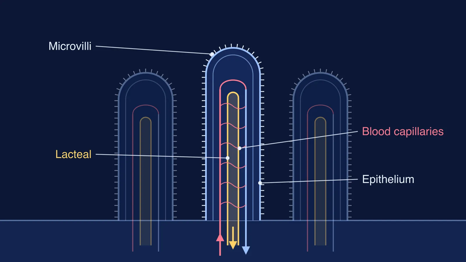
Each villus has two transport routes. OpenStax describes a capillary bed fed by one arteriole and drained by one venule, plus a lymphatic capillary called a lacteal in the centre (OpenStax).
Blood capillaries take up sugars such as glucose, and amino acids, and carry them away in the blood.
The lacteal takes up the products of fat digestion and carries them through the lymphatic system, which empties into the bloodstream.
How do you draw and label a villus for Class 10?
NCERT's Figure 5.6 shows the whole human alimentary canal, not a single villus (NCERT, p. 85), so you will usually draw the villus from memory. A clean version:
Draw a tall finger shape rising from a flat base line, rounded at the top.
Add a line just inside the outline for the lining cells, and a short fringe outside for microvilli.
Draw a narrow tube up the middle with a closed, rounded top: the lacteal.
Draw one vessel up the left side and one down the right, joined by a few wavy lines: the blood capillaries.
Add arrows at the base: blood in, blood out, lymph out.
Label checklist:
Villus
Epithelium (single layer of lining cells)
Microvilli
Blood capillaries
Arteriole (blood in) and venule (blood out)
Lacteal (lymph vessel)
Fermi's AI tutor can also draw the figure with you and check your understanding as it goes.
A three-point answer you can adapt
For "How is the small intestine designed to absorb digested food?":
The inner lining has numerous finger-like villi, which greatly increase the surface area for absorption.
The villi are richly supplied with blood vessels that carry absorbed glucose and amino acids to every cell of the body.
Each villus also has a lacteal, a lymph vessel, which absorbs fatty acids and glycerol.
Point 3 goes beyond the Class 10 NCERT text, which mentions only blood vessels; check with your teacher before relying on it.
Your next step: draw the villus once from this guide, then once from memory, and tick off the six labels.