
On this page
In 1928 a young physicist named William Astbury became a lecturer in textile physics at the University of Leeds, studying the materials fabrics are made of, such as wool and silk (Embryo Project Encyclopedia). Why does a wool fibre stretch and spring back? Astbury answered with X-rays, and showed that a protein is not a shapeless blob but a chain folded in a regular, repeating way.
Who was William Astbury?
Astbury was born on 25 February 1898 in Longton, in the Stoke district, where his father was a potter’s turner, according to the Royal Society memoir by the crystallographer J. D. Bernal (Biographical Memoirs of Fellows of the Royal Society, 1963). He won a scholarship to Jesus College, Cambridge, and worked in Leeds for thirty-three years until his death on 4 June 1961. Bernal called him “a founder of what is now one of the most exciting growing points of science, molecular biology.”
Before Leeds, Astbury learned X-ray crystallography under William Henry Bragg in London, at University College London and then the Royal Institution (William Astbury, Wikipedia). X-rays reflected from layers of atoms reveal the spacing between them; our piece on William Lawrence Bragg and the X-ray law explains how.
What did Astbury’s X-ray photographs of wool show?
A wool fibre is not a perfect crystal, but its protein, keratin, is ordered enough to scatter X-rays into a pattern of spots and arcs on film. In the first paper of their series, Astbury and A. Street reported taking “over a hundred X-ray photographs” of hair, wool and related fibres (Astbury and Street, Philosophical Transactions A, 1931).
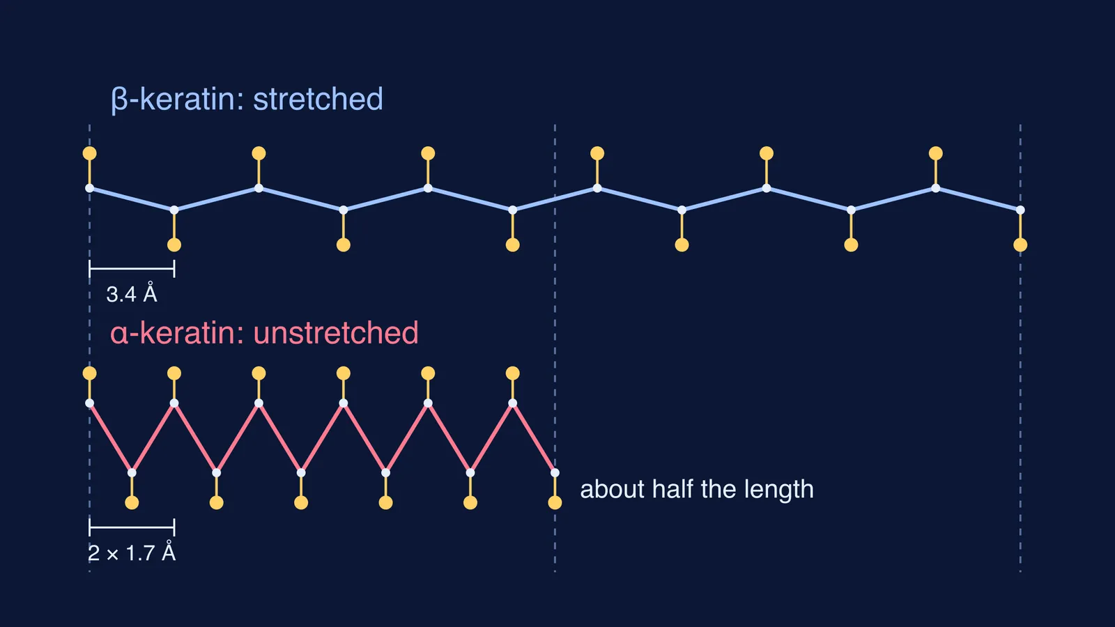
The key comparison was unstretched versus stretched. The diffraction pattern of moist wool or hair changed drastically when the fibre was stretched by about 100%, that is, to roughly twice its length (William Astbury, Wikipedia). Astbury called the normal state α-keratin and the stretched state β-keratin. A second paper, with H. J. Woods, set out to “lay bare the general structural principles underlying the properties of the protein, keratin” (Astbury and Woods, Philosophical Transactions A, 1933).
The third paper, with Wayne Sisson, summed up the model (Astbury and Sisson, Proceedings of the Royal Society A, 1935):
β-keratin is a flat “polypeptide grid” of fully extended chains bound side by side.
α-keratin is the same chains with “a regular folding of the main-chains”, which reduces the length of the molecule to approximately one-half.
Along the chain, side-chains sit about 1.7 Å apart in the folded form, against a little under 3.4 Å in the stretched form.
That is wool’s stretch in one picture: pull the fibre and you unfold the chains; let go and they fold back.
Was Astbury right about the alpha helix?
Partly: his folded model was wrong in detail, but the idea survived. In 1951 Linus Pauling, Robert Corey and Herman Branson published hydrogen-bonded helical configurations of the polypeptide chain (Pauling, Corey and Branson, PNAS, 1951). The α-helix has about 3.6 amino acid residues per turn and rises 1.5 Å per residue (Alpha helix, Wikipedia). The α and β labels in your textbook are Astbury’s names for the two forms of keratin, kept when the modern structures were worked out; read how Linus Pauling worked out the alpha helix for the rest of the story.
How much longer is a stretched protein chain?
Try this in your notebook with a keratin chain 100 amino acids long, using Astbury’s spacings (Astbury and Sisson, 1935) and the modern helix rise (Alpha helix, Wikipedia).
Stretched (β, Astbury’s spacing of about 3.4 Å): 100 × 3.4 Å = 340 Å = 34 nm.
Folded (α, Astbury’s 1935 model, 1.7 Å): 100 × 1.7 Å = 170 Å = 17 nm, exactly half.
Modern α-helix (1.5 Å per residue): 100 × 1.5 Å = 150 Å = 15 nm.
Ratio: 340 ÷ 150 ≈ 2.3, so unfolding a real helix more than doubles its length, in line with wool stretching to about twice its length.
When you revise secondary structure, fit the α-helix and β-sheet into the bigger picture with our guide to the levels of protein structure.
What can you copy from the way Astbury worked?
Start from an everyday puzzle: why does wool spring back?
Change one thing, measure again: unstretched versus stretched.
Turn pictures into numbers: 3.4 Å and 1.7 Å made the model testable.
Hold your model loosely: Astbury’s α-fold was replaced, but his names stayed.
If the jump from spots on film to spacing between atoms still feels abstract, the Fermi Simulation Lab has interactive simulations for ideas that are easier to see than to read. The Fermi Concept Library gives each concept in physics, chemistry and biology its own lesson.
Next time you pull a woollen sweater out of shape, you are doing Astbury’s experiment by hand.