On this page
In the spring of 1948, Linus Pauling was confined to his bed in Oxford with a cold. To pass the time, he began sketching polypeptide chains on a piece of paper. By folding the paper to mimic chemical bonds, he stumbled upon a corkscrew shape that satisfied all the known rules of physical chemistry.
Three years later, in April 1951, Linus Pauling, Robert Corey and Herman Branson published their formal discovery in the *Proceedings of the National Academy of Sciences* (PNAS). They had discovered the alpha helix, the fundamental structural element of proteins. A month later, Pauling and Corey published a second paper in PNAS describing the beta pleated sheet.
Together, these discoveries formed the basis of protein secondary structure, earning Pauling the 1954 Nobel Prize in Chemistry and influencing the discovery of the DNA double helix.
What is the alpha helix protein structure?
An alpha helix is a right-handed coiled sequence of amino acids. The backbone of the protein chain twists into a spiral, while the side chains (the R-groups) point outward from the core to avoid clashing with one another.
The structure is held together by hydrogen bonds. Specifically, the carbonyl oxygen (C=O) of one amino acid forms a hydrogen bond with the amide hydrogen (N-H) of the complementary group in the third amide group removed from it along the chain (PNAS). These weak but numerous bonds lock the coil into a stable shape.
The defining metric of this shape is its density. The alpha helix has 3.7 amino acid residues per turn (PNAS). This means the helix does not complete a full turn in a whole number of amino acids, a mathematical reality that had confused earlier scientists.
If you find it hard to picture how these bonds twist in 3D, the Fermi Simulation Lab has interactive simulations for ideas easier to see than read.
How did Linus Pauling discover the alpha helix in 1951?
Pauling succeeded where others failed because he refused to compromise on the rules of chemical bonds. His approach relied on two strict constraints derived from his earlier work on covalent bonds (a topic you might recognize from electron dot structures of covalent molecules in Class 10) and resonance.
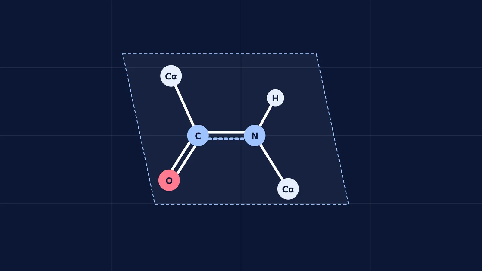
The peptide bond is planar. Because of resonance between the carbonyl carbon and the nitrogen atom, the peptide bond has partial double-bond character. This prevents rotation, meaning the Cα-C-N-Cα atoms must lie flat in a single rigid plane.
Hydrogen bonds must be linear. For maximum stability, the N-H···O atoms should lie in a nearly straight line.
Previous researchers, such as William Astbury, assumed that protein helices must have an integer number of residues per turn, such as 3 or 4. When Pauling folded his paper in 1948, he discarded the integer rule and followed the chemistry. By enforcing the planar peptide bond and straight hydrogen bonds, the 3.7 residues per turn emerged naturally (PNAS).
What is the beta pleated sheet?
In May 1951, Pauling and Corey introduced the second major piece of protein architecture: the beta sheet (PNAS).
While the alpha helix coils a single continuous chain, the beta sheet aligns multiple segments of a polypeptide chain side-by-side. The structure is held together by hydrogen bonds that form laterally between the carbonyl oxygen of one chain and the amide hydrogen of the adjacent chain.
To maintain the required bond angles and the rigidity of the planar peptide bond, the backbone cannot lie perfectly flat. Instead, it forms a zig-zag or "pleated" geometry. This pleating allows the side chains to point alternately above and below the plane of the sheet, minimizing steric hindrance.
You can review the geometry of resonance and partial double bonds in the Fermi Concept Library to see why these backbones cannot bend freely.
Why does 3.7 residues per turn matter?
The discovery of the 3.7-residue helix (PNAS) proved the value of the model-building method. By prioritizing exact bond lengths and angles over elegant mathematical integers, Pauling demonstrated that biological structures must obey the laws of physical chemistry.
This exact approach—using physical models to find shapes that satisfy chemical constraints—became the blueprint for molecular biology. Just two years later, James Watson and Francis Crick would use Pauling's model-building techniques to crack the structure of DNA.
Whether you are studying the enzymes that transcribe DNA, like the three RNA polymerases in eukaryotes, or basic biochemistry, the legacy of the 1951 papers remains. They proved that the complex machinery of life is built on simple, predictable chemical rules.