
On this page
In March 1882, Robert Koch showed that a rod-shaped bacterium causes tuberculosis. But his stain needed an hour in hot methylene blue and showed the bacilli blue on a brown background. Within 16 months, other scientists had reworked it into a recipe that turns TB bacteria bright red against a blue background. Two of them, Franz Ziehl and Friedrich Neelsen, gave the method its name. Labs still use it today, including across India, which carries a quarter of the world's TB cases according to the WHO tuberculosis fact sheet.
Who were Franz Ziehl and Friedrich Neelsen?
Franz Ziehl (1857 to 1926) was a German bacteriologist born in Wismar who held a professorship in Lübeck, according to his biography on Wikipedia. He introduced carbol fuchsin, a red dye dissolved in carbolic acid, for staining the tubercle bacillus in 1882.
Friedrich Neelsen (1854 to 1894) was a German pathologist. He was a professor at the Institute of Pathology at the University of Rostock and later worked at the pathological institute in Dresden, as his Wikipedia biography records. Their names are joined on the stain, but several people built it in stages.
How was the Ziehl-Neelsen stain developed, step by step?
A 2017 review in Microbiology Spectrum by Catherine Vilchèze and Laurent Kremer, Acid-fast positive and acid-fast negative Mycobacterium tuberculosis: the Koch paradox, lays out the timeline:
24 March 1882: Koch dipped his slides in a hot, alkaline methylene blue solution for 1 hour, then covered them with a brown dye called vesuvin.
1 May 1882: Paul Ehrlich used fuchsin or methylene violet in water saturated with aniline oil, then washed the slide with 30% nitric acid.
12 August 1882: Ziehl replaced the aniline oil with carbolic acid (phenol) and used a weaker acid wash, giving a more stable dye solution.
14 July 1883: Neelsen published a 0.75% fuchsin solution in 5% carbolic acid, decolourised with 25% sulfuric acid.
The review notes it took only 16 months to standardise the method, and that it became widely known as ZN staining about a decade later. It credits five researchers: Koch, Ehrlich, Ziehl, Rudolf Rindfleisch (who proposed heating the slides in October 1882) and Neelsen. For Ehrlich's side, read about Paul Ehrlich and his dyes, antibodies and magic bullets. For Koch's own work, see Robert Koch, tuberculosis and Koch's postulates.
Why do TB bacteria stay red after an acid wash?
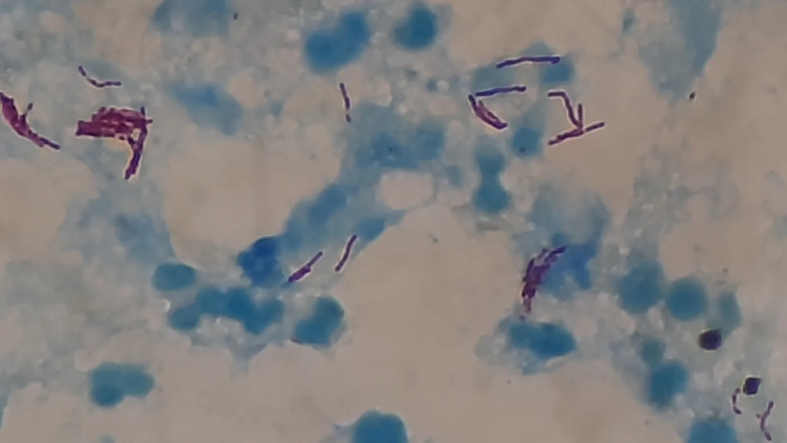
In a typical lab version, described on the teaching site of microbiologist Dr Sridhar Rao P.N., carbol fuchsin is heated on the slide until fumes rise (without letting it boil), then 20% sulfuric acid removes the dye, and Loeffler's methylene blue stains whatever is left colourless. TB bacilli show up as pink, slightly curved rods against blue pus and epithelial cells.
Acid-fast bacteria are bacteria that keep the red primary dye even after an acid wash, because the mycolic acids in their waxy envelope stop the dye washing out, as the encyclopedia entry on the stain explains. The Vilchèze and Kremer review points to mycolic acids and other cell-wall glycolipids as markers of acid-fastness, but says the exact components responsible are still largely unknown.
Compare Hans Christian Gram's stain, where alcohol sorts bacteria into two groups; here a harsher acid wash picks out the few that refuse to let go.
Is the Ziehl-Neelsen stain still used for TB today?
Yes, though it is no longer the first choice. The 2017 review's abstract calls acid-fast staining the most widely used TB diagnostic method. But a 2018 Scientific Reports study notes that guidelines ask experienced staff to read at least 100 microscope fields for at least 15 minutes per slide, and that routine reading gives a sensitivity of 60% to 70% compared with culture.
The WHO fact sheet now recommends rapid diagnostic tests, including molecular tests, as the first test for anyone with TB symptoms. It reports 10.7 million people fell ill with TB in 2024 and 1.23 million died.
How do you remember the stain for an exam or practical?
Predict the colour at each step. Take a sputum smear containing TB bacilli and human pus cells:
Carbol fuchsin with heat: everything turns red.
Acid wash: pus cells lose the red; TB bacilli keep it.
Methylene blue: pus cells turn blue; TB bacilli stay red.
Skip the acid wash: everything stays red, so you learn nothing.
Skip the methylene blue: the bacilli are red on a nearly colourless background, so they are hard to find.
Then run a three-line recall drill: write each reagent, its job, and the colour it leaves, with your notes closed. If the cell-wall chemistry behind it feels shaky, the Fermi Concept Library has a lesson for each concept in biology and chemistry, with its place in the map. And if you are not sure where to start a microbiology chapter, Fermi's AI tutor works out what you already know and builds a plan for the session.