
On this page
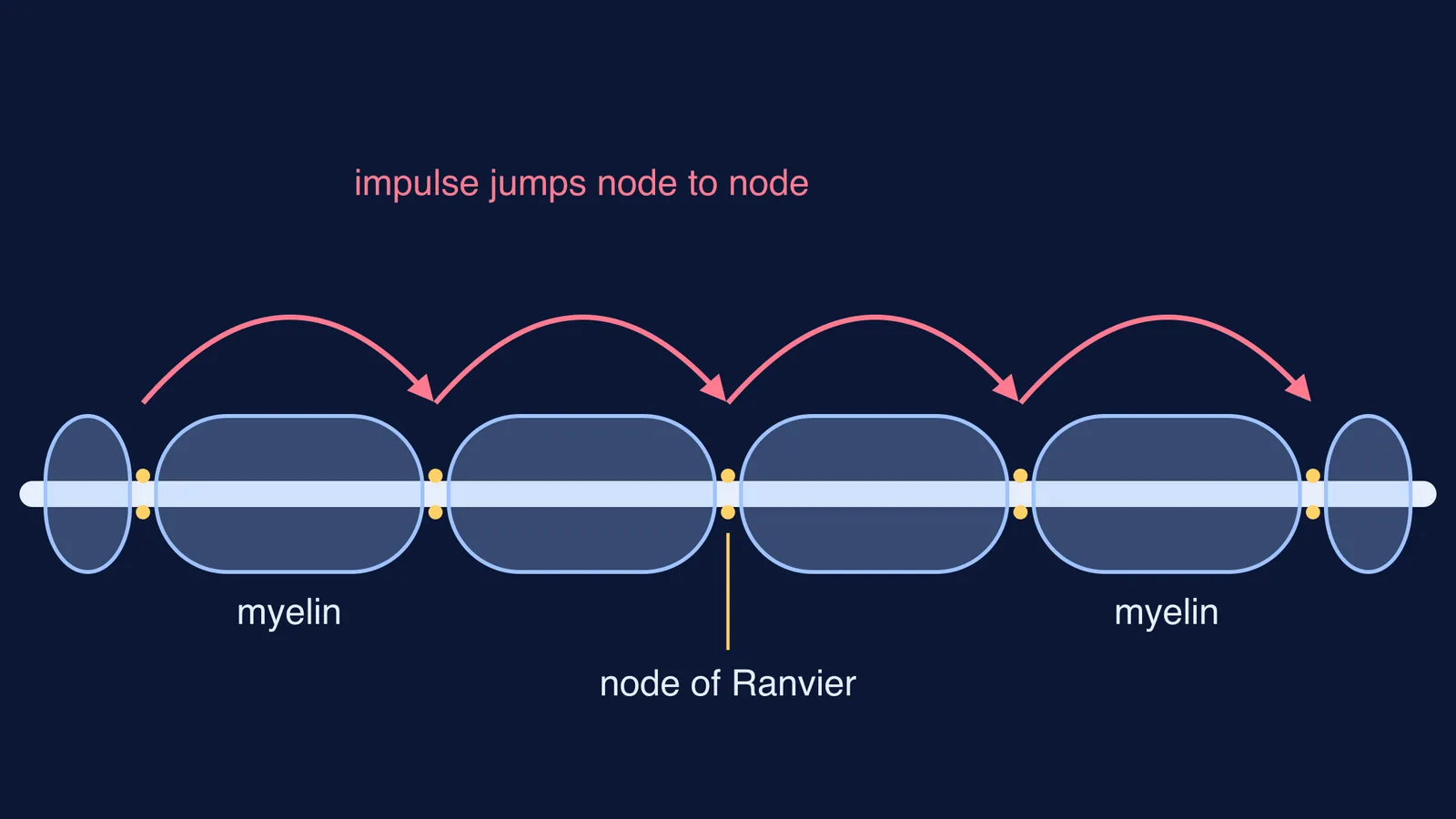
When you pull your hand back from a hot pan, the signal travels along nerve fibres, many of them wrapped in a fatty coat called myelin. That coat is not continuous. It is broken at regular points by tiny bare gaps, and those gaps carry the name of the man who first described them: Louis-Antoine Ranvier.
Who discovered the nodes of Ranvier?
Louis-Antoine Ranvier was a French histologist, born in Lyon on 2 October 1835, who held the chair of general anatomy at the Collège de France from 1875 and died in 1922 (Wikipedia biography). Histology is the study of tissues under the microscope.
A 2019 review in Arquivos de Neuro-Psiquiatria, written for the 140th anniversary of his book, credits Ranvier with the first description of the nodes (Fustes and colleagues, 2019). Working in his Paris laboratory around 1871, he treated frog sciatic nerves with osmic acid. Along each fibre he saw narrowings, or constrictions, roughly 1 mm apart, where the myelin stopped.
In 1878 he gathered this work into his *Leçons sur l'histologie du système nerveux* (Lessons on the histology of the nervous system). The same review notes that he proposed there that myelin acted as an electrical insulator, interrupted at points along the axon. Those interruptions are what we now call the nodes of Ranvier.
Why do the gaps make nerve signals faster?
Ranvier saw the structure. Proving what it did took more than 60 years. The 2019 review records that Tasaki and Takeuchi in 1941, and A. F. Huxley and R. Stämpfli in 1949, showed that in myelinated fibres the electrical change happens at the nodes, not along the insulated stretches between them (Fustes and colleagues, 2019). Huxley and Stämpfli titled their paper *Evidence for saltatory conduction in peripheral myelinated nerve fibres* (Journal of Physiology, 1949).
Here is the mechanism in plain terms. Under the myelin, there are no voltage-gated sodium channels until the next node, so the impulse cannot be regenerated there (OpenStax Anatomy and Physiology). Current spreads under the insulation, then the next node fires a fresh action potential. The signal appears to leap from node to node.
The payoff is speed. A 2015 modelling study notes that this jumping lets action potentials travel at up to 100 m/s or more (Johnson and colleagues, Journal of Neurophysiology). For a side-by-side comparison of the two fibre types, see our explainer on myelinated vs non-myelinated nerve fibres. For the action potential itself, read how Hodgkin and Huxley explained the action potential.
How small is a node of Ranvier?
Tiny. The same 2015 study states that the nodal gap is generally about 1 μm long, while the insulated internode can exceed 1 mm in large axons (Johnson and colleagues, 2015). In rat optic nerve, an eLife team measured a mean node length of 1.02 μm and modelled internodes about 139 μm long, a value taken from earlier measurements (Arancibia-Cárcamo and colleagues, eLife 2017).
Node size is not a trivial detail. The eLife team found node lengths in rat cortex varied over an 8.7-fold range and modelled that such differences could change conduction speed by about 20% (Arancibia-Cárcamo and colleagues, 2017).
Worked example: how much of an axon is bare?
Use the numbers above to see why the design is efficient.
Large axon: a node about 1 μm long every 1 mm (1,000 μm) of myelin. Bare fraction = 1 ÷ 1,001, about 0.1% of the fibre.
A 1 metre stretch of that axon has about 1,000 nodes (1 m ÷ 1 mm).
At 100 m/s, the impulse covers that metre in 1 ÷ 100 s, which is 10 milliseconds.
Rat optic nerve: 1.02 ÷ (139 + 1.02), about 0.7% of the fibre is node.
The axon regenerates the signal along less than 1% of its length.
How do you answer exam questions on the nodes of Ranvier?
Biology papers tend to test the same few points. Check yourself against this list:
Who and when: Louis-Antoine Ranvier, French histologist; described the gaps in the 1870s and published them in his 1878 lectures.
What a node is: a short gap in the myelin sheath where the axon membrane is exposed.
What happens there: the action potential is regenerated at each node; between nodes, current spreads under the myelin.
Name of the process: saltatory conduction.
Why it matters: fast conduction without needing a much thicker axon.
Common trap: jumping does not mean the signal vanishes between nodes; current spreads under the myelin and the action potential is rebuilt at the next node.
A jump from node to node is hard to picture from a textbook diagram alone. The Fermi Simulation Lab has interactive simulations for ideas that are easier to see than to read, and if a related term trips you up, the Fermi Concept Library gives each biology concept its own lesson and its place in the map.