
On this page
Ask who invented the microscope and you get two answers. Rival claims point to Dutch spectacle makers and the inventor Cornelis Drebbel, and nobody agrees who was first. And a Delft cloth merchant, Antonie van Leeuwenhoek, used a single bead of glass to describe bacteria before anyone else.
Who invented the microscope?
The history section of Wikipedia's microscope article lists rival claims: Zacharias Janssen in 1590 (a claim made by his son), his father Hans Martens, their neighbour Hans Lippershey, and Cornelis Drebbel, who was noted to have one in London in 1619. The same article says the earliest known compound microscopes, with an objective lens near the specimen and an eyepiece, appeared in Europe around 1620.
Galileo built an improved one after seeing Drebbel's exhibited in Rome in 1624, and called it the occhiolino, or “little eye” (Wikipedia). These first instruments were weak. A tube microscope attributed to Janssen magnified about three times when closed and up to ten times when extended, according to Florida State University's Molecular Expressions museum.
How did Leeuwenhoek's single-lens microscope work?
Leeuwenhoek (1632–1723) ran a draper's shop and wanted to see the quality of thread better than the magnifying lenses of the time allowed (Wikipedia). His microscopes were small, the largest about 5 cm long, each holding one tiny lens. You held the lens very close in front of your eye, and the specimen sat on a pin on the other side (Wikipedia).
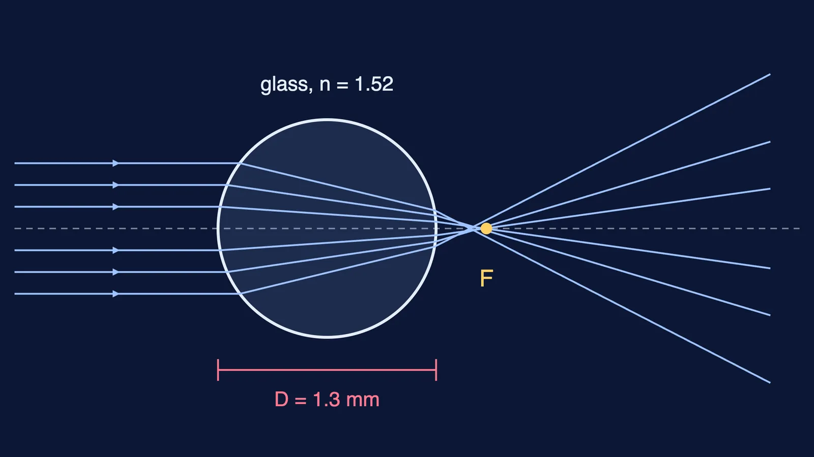
In 2021 a team from Rijksmuseum Boerhaave and TU Delft used neutron tomography to look inside two of his surviving instruments (Cocquyt et al., Science Advances; open copy). Of the hundreds of microscopes he made, they write, only a handful survive. The one with the highest magnification, at Utrecht University Museum, magnifies 266 times and has a measured resolving power of 1.35 μm. Its lens was not ground. It is a flameworked glass globule 1.3 mm across, with a tiny glass stem still attached, made by a method Robert Hooke popularised in 1678. The authors call this microscope unsurpassed in optical performance until the 1830s.
How much does a 1.3 mm glass bead magnify?
You can check the 266 measured by Cocquyt and colleagues with Class 12 ray optics.
Apply refraction at a curved surface at both sides of a sphere of diameter D and refractive index n. Parallel light focuses at f = nD / 4(n − 1), measured from the centre.
Take ordinary glass, n ≈ 1.52, and D = 1.3 mm: f = (1.52 × 1.3) / (4 × 0.52) = 1.976 / 2.08 ≈ 0.95 mm.
A simple microscope with the final image at infinity has magnifying power M = 25 cm / f, where 25 cm is the near point of a normal eye.
M = 250 mm / 0.95 mm ≈ 263.
That is within about 1% of the measured 266. His glass's exact refractive index is unknown, so treat this as a check, not a measurement. The catch: a focal length under a millimetre puts the specimen almost touching the lens, and the lens almost touching your eye. The same logic explains why a microscope objective has a short focal length and how far to hold a magnifying glass for the biggest image.
If light bending at a curved surface is hard to picture, the Fermi Simulation Lab has interactive simulations for ideas that are easier to see than to read.
What did Leeuwenhoek see first?
His first letter to the Royal Society in London, published in 1673, described mould, bees and lice (Wikipedia). The biologist Nick Lane's 2015 commentary in Philosophical Transactions B (open copy) retells what came next:
In 1675 he found living creatures in rain water that had stood a few days in a new earthen pot.
In his letter of 9 October 1676, the “letter on the protozoa”, he reported more than 1,000,000 living creatures in one drop of pepper-water. Later he wrote that he could truthfully have said eight times as many.
He judged that 100 of the smallest, laid end to end, would not reach the length of a coarse grain of sand. From his own later estimate of the grain's size, Lane puts them at less than 3 µm long: bacteria.
The Royal Society asked Nehemiah Grew to repeat the work, and he failed. Then in 1677 Robert Hooke took up microscopy again and, after early failures, saw the animalcules, and Leeuwenhoek was elected a Fellow in 1680 (Lane, 2015). Lane adds that later scientists could not match the resolution of his microscopes, and that twentieth-century work confirmed he had seen bacterial cells.
What can you take from a glass bead?
Magnifying power comes from a short focal length. With M = 25 cm / f, halving f doubles M.
More lenses do not automatically mean a better instrument: his one-lens Utrecht microscope was unsurpassed until the 1830s (Cocquyt et al.).
Write down what you measure. His sand-grain comparison could still be turned into micrometres three centuries later.
For magnification numericals in ray optics, the Fermi Problem Library has worked problems with a step-by-step solver that explains the reasoning rather than printing an answer.