
On this page
In May 1934, the journal Nature printed a short letter by J. D. Bernal and Dorothy Crowfoot titled "X-Ray Photographs of Crystalline Pepsin" (Bernal and Crowfoot, Nature, 1934). It reported X-ray photographs that showed a protein crystal as an ordered, repeating structure instead of a smudge. The trick that made it work was simple: keep the crystal wet.
Who was J. D. Bernal and what did he photograph in 1934?
Bernal was an X-ray crystallographer working in Cambridge in the early 1930s. Dorothy Crowfoot, later Dorothy Hodgkin, came from Oxford to work with him and spent two years there, according to her Nobel Prize biography.
The crystals were pepsin, the protein-digesting enzyme of the stomach. The Nature letter says Dr. G. Millikan brought them "prepared by Dr. Philpot in the laboratory of Prof. The Svedberg, Uppsala", and that they were "perfect hexagonal bipyramids up to 2 mm. in length" (Nature, 1934).
Hodgkin later told the story on camera. Glenn Millikan saw John Philpot's crystals and said, "I know a man in Cambridge who would give his eyes for those crystals"; Philpot offered him a tubeful, which travelled back to Cambridge in a shirt pocket (Dorothy Hodgkin, Web of Stories).
Why did earlier X-ray photos of proteins show nothing?
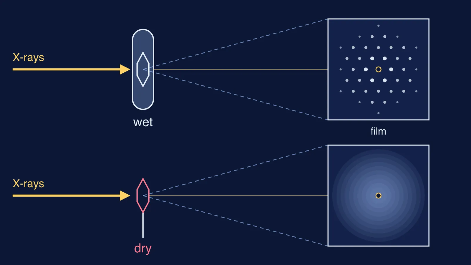
Because the crystals had dried out. In their own liquid the pepsin crystals looked bright and orderly under polarised light, but "on exposure to air, however, the birefringence rapidly diminishes", Bernal and Crowfoot wrote (Nature, 1934).
Photographs "taken of the crystals in the usual way showed nothing but a vague blackening". The letter concluded that this showed "complete alteration of the crystal" and "explains why previous workers have obtained negative results with proteins, so far as crystalline pattern is concerned".
Hodgkin described the moment in her Web of Stories interview. Bernal raked a crystal out of the tube with a needle and mounted it on a glass fibre, his usual method. It looked "a bit shrivelled", and the X-rays gave "almost nothing". He reasoned that the crystal no longer had water around it, borrowed Lindemann glass tubes from his research student Helen Megaw, and sealed a crystal inside with its mother liquor, the solution it grew in.
The next photograph, Hodgkin recalled, "was full of reflections, rather large, blotchy reflections". She added that the original film "seems to have disappeared since".
What did the wet pepsin photograph prove?
X-rays scattered by a regular, repeating arrangement of atoms add up in a few directions and make spots; a jumbled arrangement spreads them into a blur. So spots from wet pepsin meant a protein could form a truly ordered crystal, as long as its water stayed with it. The Complete Dictionary of Scientific Biography calls it "the first demonstration of atomic regularity in a globular protein" (Encyclopedia.com).
That opened the door to reading a protein's shape from its spots. Crowfoot went on to win the 1964 Nobel Prize in Chemistry "for her determinations by X-ray techniques of the structures of important biochemical substances" (Nobel Prize); read how Dorothy Hodgkin used X-rays to reveal penicillin, B12 and insulin. For the first complete protein structure, read how John Kendrew solved myoglobin.
How can you use Bernal's method when your own experiment fails?
Bernal's first try failed, and the way he recovered is a method you can copy in a school lab or a physics numerical.
Write down exactly what you saw. Bernal's record was "a vague blackening", not "it didn't work".
Ask what changed between the thing in its natural state and the thing in your test. The pepsin left its liquid and shrivelled.
Change one condition only. Same crystals, same X-rays; only the water came back.
Compare the new result with the old one side by side, and keep both.
Here is a worked example from Class 10 chemistry. You heat blue copper sulphate crystals and they crumble into a white powder. Step 1: note "blue crystals became white powder, and droplets appeared at the cool end of the tube". Step 2: what left? Water. Step 3: add a few drops of water to the cooled powder, changing nothing else. Step 4: it turns blue again. Like Bernal's pepsin, the water was part of the crystal. To put numbers on it, see how to find the water of crystallisation in a hydrate from mass loss.
If the physics is what stops you, why order gives spots and disorder gives a blur, it can help to see it rather than read it. The Fermi Simulation Lab has interactive simulations for ideas that are easier to see than to read, and the Fermi Concept Library gives every concept in physics, chemistry and biology its own lesson, so you can fill the gap before the next practical.
The next time a result comes back as a blur, ask Bernal's question first: what did my sample lose on the way to the test?