On this page
In March 1958, John Kendrew and five colleagues published "A three-dimensional model of the myoglobin molecule obtained by X-ray analysis" in Nature 181, 662-666. Within two years, the same team had sharpened the picture enough to build an atomic model. Myoglobin was the first protein to have its atomic structure determined, as the Protein Data Bank's PDB-101 Molecule of the Month notes.
Who was John Kendrew and why did he choose myoglobin?
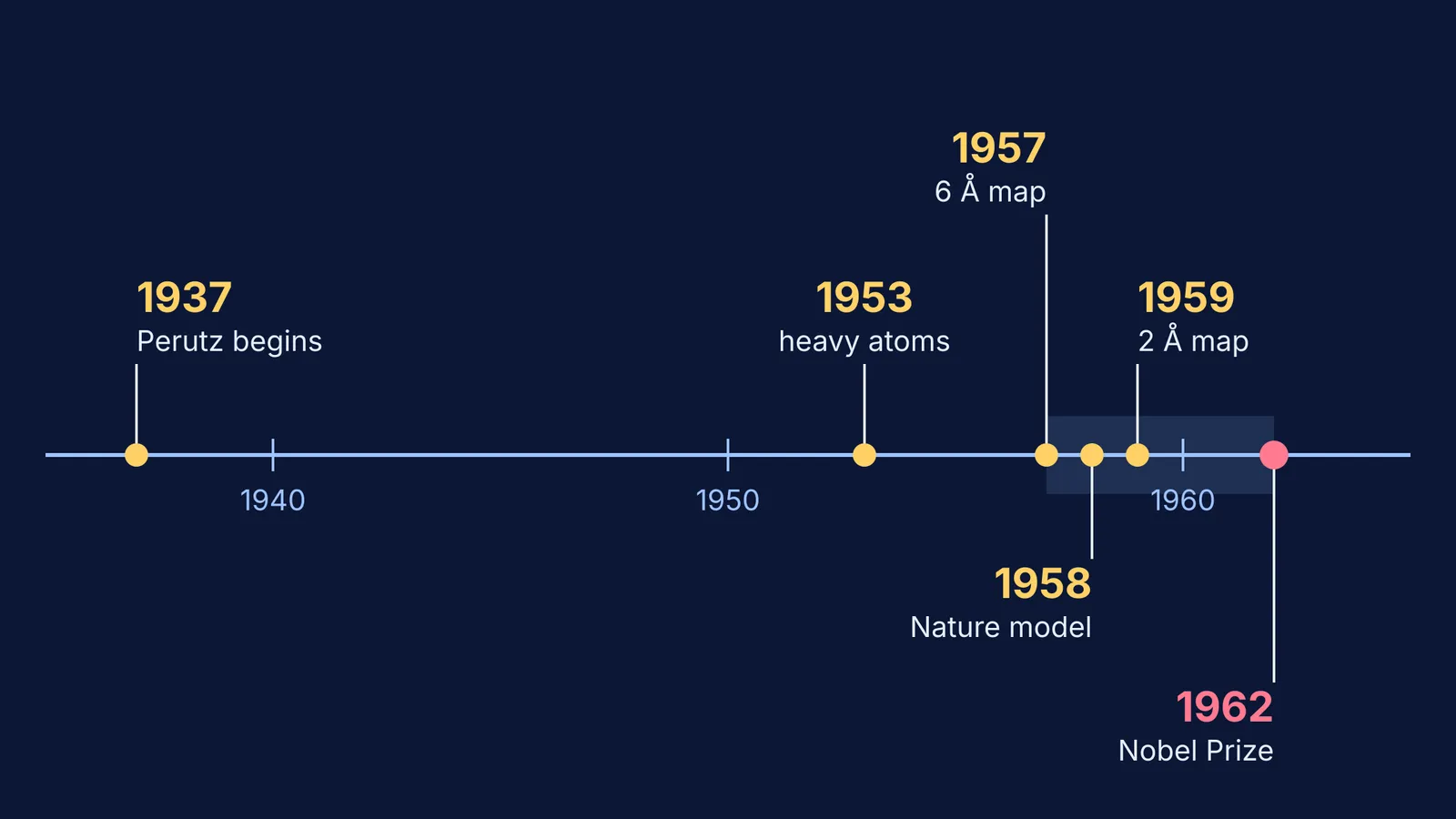
Kendrew was born in Oxford in 1917, as Max Perutz recalled in his obituary of Kendrew for the British Crystallographic Association. According to the 1962 Nobel presentation speech, Perutz began X-ray experiments on haemoglobin in 1937. About ten years later Kendrew joined his group and was given myoglobin.
Myoglobin stores oxygen in muscle. It is closely related to haemoglobin but only a quarter as large, the speech notes. Perutz wrote that this made it "a more hopeful candidate for X-ray study". Horse heart myoglobin would not give large enough crystals, so Kendrew turned to diving mammals and birds, in which nearly one tenth of the dry weight of the muscles is myoglobin. A chance encounter let Perutz get him "a large chunk of sperm whale meat from Peru". For the two proteins side by side, see our explainer on haemoglobin vs myoglobin.
How did X-ray crystallography reveal myoglobin's structure?
When an X-ray beam meets a crystal, it sends out beams called reflections, and their directions and intensities depend on the type and arrangement of the atoms, as the Nobel presentation speech explains.
The catch is called the phase problem. The diffraction pattern records the amplitudes of the diffracted rays but not their phases, and you need both to rebuild the molecule. The answer was to compare the diffraction pattern of a plain protein crystal with that of a crystal carrying heavy atoms, as Perutz described in his obituary of Kendrew. Perutz did this with mercury in haemoglobin in 1953. Kendrew found another way to attach heavy atoms, usually mercury or gold, to myoglobin.
For this to work, the heavy atoms must not move the rest of the protein. The speech quotes Lawrence Bragg: "the molecule takes no more notice of such an insignificant attachment than a maharaja's elephant would of the gold star painted on its forehead". You can read more about the trick in our post on Max Perutz and the structure of haemoglobin.
Was the 1958 myoglobin model atom by atom?
Not yet. By 1957 the team had an electron density map at 6 Å resolution. That was enough for a rough molecular model. Two years later they reached 2.0 Å, which let Kendrew build an atomic model, "the first of any protein", according to Perutz's obituary. The sharper map was published in 1960 as "Structure of myoglobin: a three-dimensional Fourier synthesis at 2 Å resolution" in Nature 185, 422-427.
The numbers show how big the job was. Myoglobin contains about 2,600 atoms, and Kendrew examined 110 crystals and measured the intensities of about 250,000 X-ray reflections, according to the Nobel presentation speech.
Kendrew and Perutz shared the 1962 Nobel Prize in Chemistry "for their studies of the structures of globular proteins" (Nobel Prize summary). A later refinement of the sperm whale myoglobin model by Watson and Kendrew, at 2.0 Å, is entry 1MBN in the Protein Data Bank.
What do Kendrew's numbers tell you? A worked example
Take the three figures from the Nobel presentation speech: 2,600 atoms, 110 crystals, 250,000 reflections.
Reflections per crystal: 250,000 / 110 is about 2,300 on average.
Measurements per atom: 250,000 / 2,600 is about 96. Each atom has three unknown coordinates (x, y, z), so that is roughly 30 measurements for every unknown. Many came from the heavy-atom crystals used for phasing, but far more measurements than unknowns is what pins a structure down.
Scaling up: the speech says haemoglobin is four times as large, so roughly 10,000 atoms. In 1962 its structure was known less thoroughly than myoglobin's.
What can students learn from how Kendrew worked?
His method doubles as a way to study a hard chapter:
Start with the smaller case. He picked myoglobin, a quarter the size of haemoglobin. Learn one clean example well before the messy one.
Get the rough shape first. He went from 6 Å to 2 Å. Sketch the chapter's big picture, then fill in the detail.
Use contrast. The heavy-atom trick compared two almost identical crystals. Comparing two similar ideas makes each easier to remember.
Know the levels. "Structure" covers primary to quaternary. Our guide to the four levels of protein structure shows where myoglobin's folded chain fits.
If an idea is hard to picture from a paragraph, the Fermi Simulation Lab has interactive simulations for ideas that are easier to see than to read. If one term is the gap, the Fermi Concept Library gives each concept its own lesson.
The first protein structure came from whale muscle, heavy atoms and about 250,000 measured reflections.