

On this page
Every breath you take sends oxygen into your bloodstream, where it meets its first carrier: haemoglobin. But by the time that oxygen reaches your muscles, it needs a different molecule to take over the load. That molecule is myoglobin.
While both are iron-rich, heme-containing proteins built on a similar folding pattern, they do entirely different jobs. Haemoglobin is built for transport, meaning it has to be good at both picking up oxygen and letting it go. Myoglobin is built for storage, meaning it holds on tight until the cell is desperate.
What is the structural difference between haemoglobin and myoglobin?
The fundamental difference lies in their size and complexity.
Haemoglobin is a tetramer. It is made of four polypeptide chains (two alpha and two beta chains in adults). Because it has four subunits, it contains four heme groups and can bind up to four molecules of oxygen at once. You will find it packed inside your Formed elements of blood, specifically the red blood cells.
Myoglobin is a monomer. It consists of a single polypeptide chain with just one heme group, meaning it can only bind one molecule of oxygen. It is found primarily in muscle tissue.
This difference in structure is the reason why red muscle fibres—also called slow-twitch or Type I fibres—are red. These fibres rely heavily on sustained aerobic respiration (a process related to the Respiratory quotient of the fuels they burn). To keep oxidative phosphorylation going without fatigue, they pack in a dense supply of myoglobin, which gives the meat its dark, rich colour.
Why does myoglobin have a higher oxygen affinity than haemoglobin?
The job of myoglobin is to act as a stockpile. To do this, it must grab oxygen out of the blood and refuse to let it go until the muscle absolutely needs it.
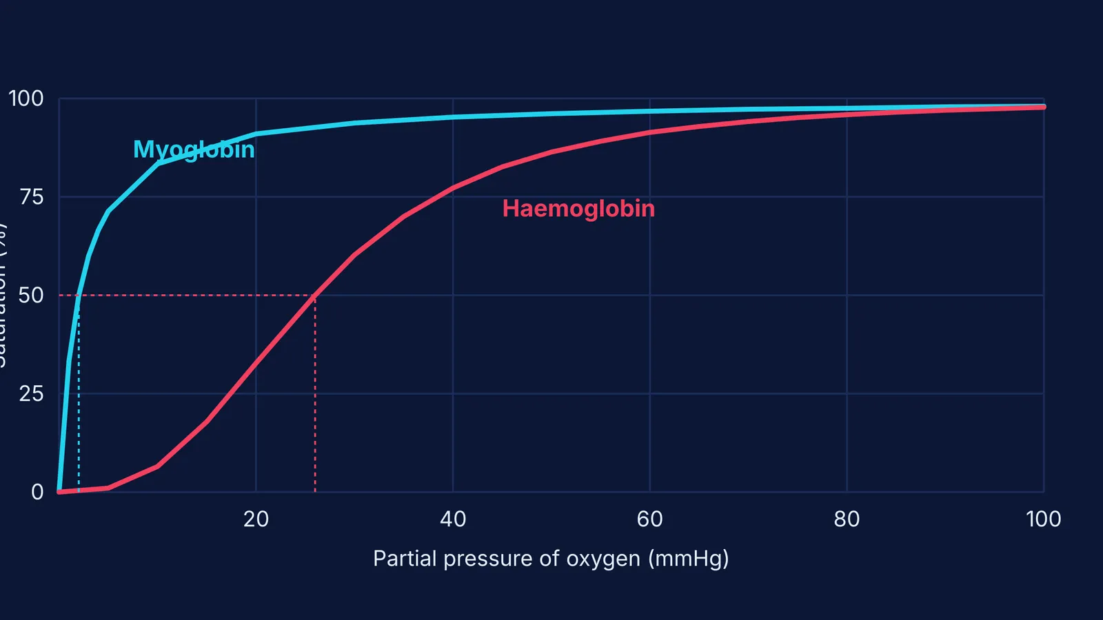
Biochemists measure this grip using the P₅₀ value: the partial pressure of oxygen at which a protein is exactly 50% saturated. A lower P₅₀ means a stronger grip. According to Biochemistry (5th edition), the P₅₀ of myoglobin is roughly 2 mmHg, while the P₅₀ of haemoglobin is much higher, at approximately 26 mmHg.
Because myoglobin's affinity is so high, it remains almost fully saturated at the oxygen pressures where haemoglobin is actively unloading. Only when the muscle contracts intensely and cellular oxygen levels plummet does myoglobin finally release its reserve to the mitochondria.
How do their oxygen dissociation curves compare?
If you graph how much oxygen these proteins hold at different pressures, the structural difference becomes visible.
Myoglobin binds oxygen independently because it only has one subunit. Its graph is a simple hyperbolic curve. It shoots up quickly at very low oxygen pressures and flattens out near 100% saturation.
Haemoglobin, on the other hand, shows cooperative binding. When one of its four subunits binds to an oxygen molecule, it changes the shape of the entire tetramer, making it easier for the other three subunits to bind oxygen. This creates a sigmoidal (S-shaped) curve.
This S-shape is perfect for a transport protein. In the lungs, where oxygen pressure is high, haemoglobin loads up fully. In the tissues, where the pressure drops, the curve dips steeply, allowing haemoglobin to dump a large fraction of its oxygen payload right where it is needed.
If you keep mixing up transport and storage proteins, the Fermi Concept Library maps out respiratory physiology so you can see exactly how the curves and structures connect.
How does structure dictate cooperative binding?
The tetrameric structure of haemoglobin shifts between two distinct forms: the T (tense) state and the R (relaxed) state. In the T state, subunits are tightly bound together by salt bridges, leaving a low affinity for oxygen.
When the first oxygen binds to a heme group, it pulls the iron atom into the porphyrin ring. This tiny movement breaks the salt bridges and triggers a rotation between the dimers. This shift forces the entire tetramer into the R state, drastically raising its oxygen affinity. Binding the first oxygen makes it easier for the remaining sites to fill.
The crucial role of 2,3-BPG
Pure haemoglobin holds onto oxygen too tightly to be a good transport protein. It requires an allosteric effector called 2,3-bisphosphoglycerate (2,3-BPG) to function in your red blood cells.
2,3-BPG slips into the centre pocket of the haemoglobin tetramer, but only fits when the protein is in the T state. By binding to positively charged residues, it stabilizes the tense conformation. This lowers oxygen affinity, ensuring oxygen is released into metabolizing tissues. In the lungs, high oxygen forces the R state, which collapses the pocket and squeezes 2,3-BPG out.
How does the Bohr effect separate them further?
Because haemoglobin is a complex tetramer, it can be regulated by its environment—a phenomenon known as allosteric regulation.
When a muscle works hard, it produces carbon dioxide and hydrogen ions, lowering the local pH. This acidic environment shifts haemoglobin's dissociation curve to the right (the Bohr effect), forcing it to unload more oxygen. At a molecular level, the lower pH protonates specific amino acids, allowing them to form salt bridges that lock the protein into the T state. Carbon dioxide also binds directly to the terminal amino groups to form carbamates, further stabilizing this state.
Myoglobin, being a simple monomer, is blind to these changes. It ignores the Bohr effect entirely, steadfastly holding its oxygen until the pressure drops low enough to physically pull it away. This division of labour ensures that oxygen always flows smoothly from the lungs, through the blood, and into the working engine of the cell.
A quick framework for exam questions
If you face a multiple-choice question on respiratory pigments in an exam, use this three-point checklist to find the right answer immediately:
Check the location: Is the question asking about the bloodstream or the muscle? Blood means haemoglobin; muscle means myoglobin.
Check the shape: Does the prompt mention an S-shape or cooperativity? A sigmoidal curve always belongs to the tetramer (haemoglobin).
Check the shift: Is the protein reacting to an acidic environment or high carbon dioxide (the Bohr effect)? Only haemoglobin responds to these allosteric triggers.


