
On this page
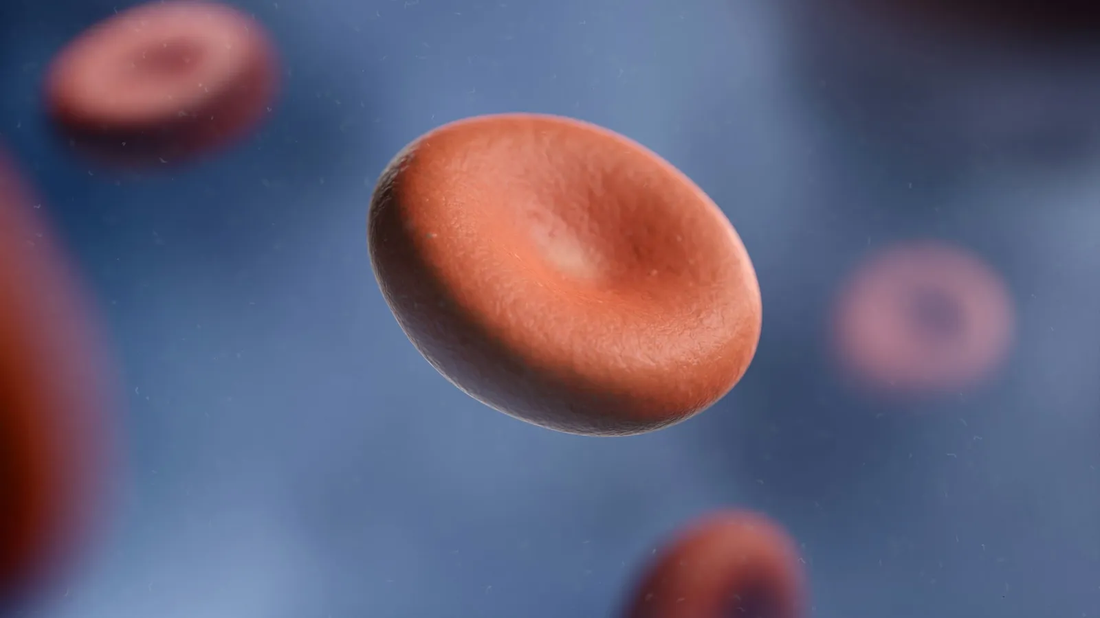
NCERT tells you that each haemoglobin molecule can carry at most four molecules of oxygen. Max Perutz spent more than twenty years finding out what that molecule looks like, and his methods opened the door to seeing the shapes of proteins in general. This post covers his life, how the structure was solved, a worked example using his numbers, the usual mistakes, and what students can learn from his persistence.
Who was Max Perutz?
His Nobel facts page gives his birth in Vienna on 19 May 1914 and his death in Cambridge on 6 February 2002. His Nobel biography says his parents wanted him to study law and join the family textile business, but a good schoolmaster turned him towards chemistry. He joined the Cavendish Laboratory in Cambridge as a research student under J. D. Bernal in September 1936.
When Austria was annexed, the family business was taken, his parents became refugees and his money ran out. On 1 January 1939 he was appointed research assistant to Sir Lawrence Bragg on a Rockefeller Foundation grant. In 1947 he became head of a new Medical Research Council unit, with John Kendrew as its entire staff. That unit grew into the MRC Laboratory of Molecular Biology.
He shared the 1962 Nobel Prize in Chemistry with Kendrew "for their studies of the structures of globular proteins".
Why was haemoglobin so hard?
The biography says the work began after a conversation in Prague in September 1937. G. S. Adair made him the first crystals of horse haemoglobin. In his Nobel lecture Perutz explains the obstacle. Shine X-rays on a crystal and you get a pattern of spots. Each spot gives the strength of one wave, but not its phase, the position of its crests. "By itself, the X-ray pattern tells only about the amplitude, but not about the phase angles," so "half the information needed for production of the image is missing."
How did heavy atoms solve it?
His answer was isomorphous replacement: make crystals that are identical except for a heavy atom, such as mercury, fixed at one spot on each protein molecule. The heavy atom changes the spot intensities slightly, and comparing the two patterns reveals the phases. A second heavy-atom crystal removes the remaining ambiguity.
In 1954, with D. W. Green and V. M. Ingram, he showed in the Proceedings of the Royal Society that crystals of horse haemoglobin with mercury compounds attached were "exactly isomorphous" with normal ones, yet changed the intensities of many reflections. Using them, the signs of just over two-thirds of the reflections in one projection could be found with certainty.
The full 3D map came in 1959 and was published in Nature in 1960 at 5.5 Å resolution. The Nobel lecture notes that a related myoglobin calculation needed about a quarter of a million measured spots and about 5 × 10⁹ additions and subtractions, impossible without the new electronic computers.
What did the map show?
The lecture describes a molecule of molecular weight 64,500 with about 10,000 atoms, four of them iron. There are four haem groups and four chains, identical in pairs, each with just over 140 amino acids. The biggest surprise was that the haem groups sit far apart, in separate pockets on the surface, even though binding oxygen at one makes the others bind more readily. Comparing oxygenated horse haemoglobin with oxygen-free human haemoglobin, he found the two β chains moved apart by more than 7 Å. In 1970 he published his explanation of how the molecule switches, in Stereochemistry of cooperative effects in haemoglobin.
Worked example: how much oxygen can blood carry?
NCERT's Breathing and Exchange of Gases chapter says each haemoglobin molecule carries at most four O₂ molecules, and Body Fluids and Circulation says a healthy person has 12 to 16 g of haemoglobin per 100 mL of blood.
Moles in 1 g. 1 g ÷ 64,500 g/mol = 1.55 × 10⁻⁵ mol of haemoglobin.
Oxygen it can hold. × 4 = 6.20 × 10⁻⁵ mol of O₂.
As a gas volume. At STP, 1 mol occupies 22.4 L, so 6.20 × 10⁻⁵ × 22,400 mL = 1.39 mL of O₂ per gram.
Per 100 mL of blood. With 15 g of haemoglobin, 15 × 1.39 = 20.8 mL of O₂ when fully saturated. The NCERT range gives 16.7 to 22.2 mL.
NCERT says that 100 mL of oxygenated blood delivers around 5 mL of O₂ to tissues at rest. That is about a quarter of the 20.8 mL carried, so most oxygen comes back to the lungs still bound.
What mistakes do students make here?
Four, not one. One haemoglobin molecule binds up to four O₂, one per haem iron.
Iron stays Fe²⁺. The lecture notes that the iron combines with oxygen "without itself becoming oxidized".
Carbon dioxide is not carried on the iron. Perutz's lecture says CO₂ is not bound by the iron atoms directly.
Molecular mass of the molecule, not one chain. Use 64,500 for the whole tetramer.
Our post on breathing and gas exchange covers the dissociation curve, and Dorothy Hodgkin covers the X-ray method from another angle. If the sigmoid curve is hard to picture, the Fermi Simulation Lab has interactive simulations for ideas easier to see than read.
What can you take from how he worked?
Stay with a hard problem. He began in 1937 and saw the structure in 1959.
Change the method when it stalls. The heavy-atom trick turned an impossible calculation into a hard one.
Cross subject lines. The biography describes him cycling between Keilin's biochemistry lab and the Cavendish, "bridging the gap between biology and physics on his bicycle".
Next step: redo the worked example for a person with 12 g of haemoglobin per 100 mL and compare it with the 5 mL delivered at rest.