
On this page
In 1904 a chemist in Hamburg published a small change to a blood stain: he added glycerol so the dye solution stayed stable. That chemist was Gustav Giemsa, and his stain is still how laboratories look for malaria parasites in a drop of blood. Here is what he actually fixed, and how to read a stained smear yourself.
Who invented the Giemsa stain?
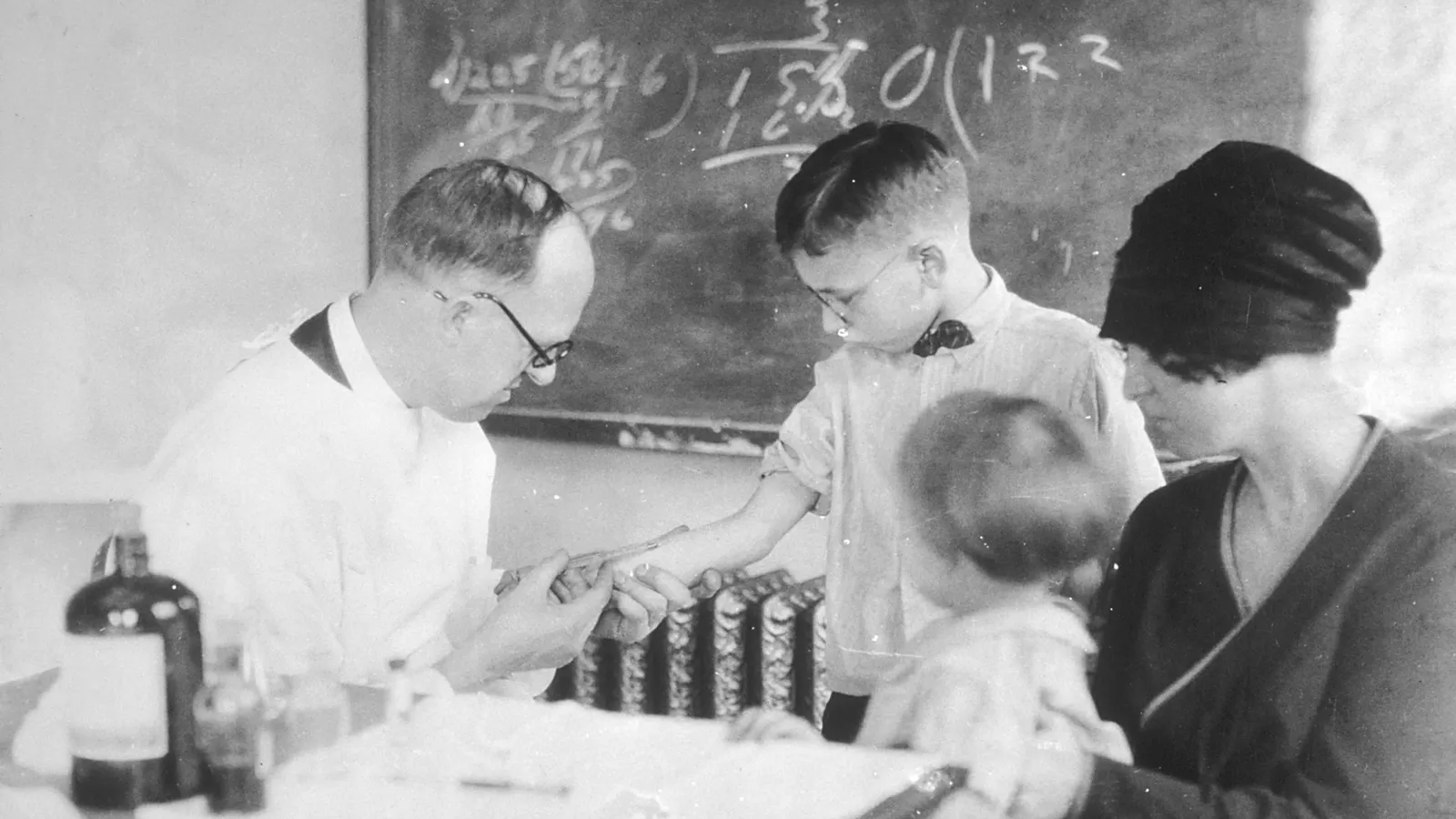
Gustav Giemsa was born on 20 November 1867 in Blechhammer, Upper Silesia, and died on 10 June 1948 in Biberwier, Tyrol, according to the Deutsche Biographie. He studied pharmacy in Leipzig from 1892 to 1894, and later organic and physiological chemistry at the University of Berlin. In 1900 he took charge of the chemistry department at Bernhard Nocht's Institute for Maritime and Tropical Diseases in Hamburg.
He did not start from nothing. In 1891 the Russian physician Dmitri Romanowsky developed a stain from eosin and aged solutions of methylene blue and used it on blood infected with malaria (Romanowsky stain overview). The trouble was consistency. The "polychromed" methylene blue used in these recipes could contain up to 11 different dyes, and even batches from the same maker varied.
Giemsa's answer came in two steps, both published in the *Zentralblatt für Bakteriologie*: a formula built on a methylene-azure mixture in 1902 (Deutsche Biographie), and in 1904 the addition of glycerol to make the solution more stable (Romanowsky stain overview; Gustav Giemsa biography). So the honest answer to "who invented Giemsa stain" is this: Giemsa made Romanowsky's idea reliable enough to use every day.
The parasite itself had been seen years earlier, as our story on Alphonse Laveran and the discovery of the malaria parasite tells. Giemsa's contribution was making it easy to find.
How does Giemsa stain show malaria parasites?
A pathologist's history of the stain, Barcia (2007) in the International Journal of Surgical Pathology, says the use of methylene azure, mixed with methylene blue to form an eosinate, is what made the stain and its results stable. It was designed mainly to show malaria parasites.
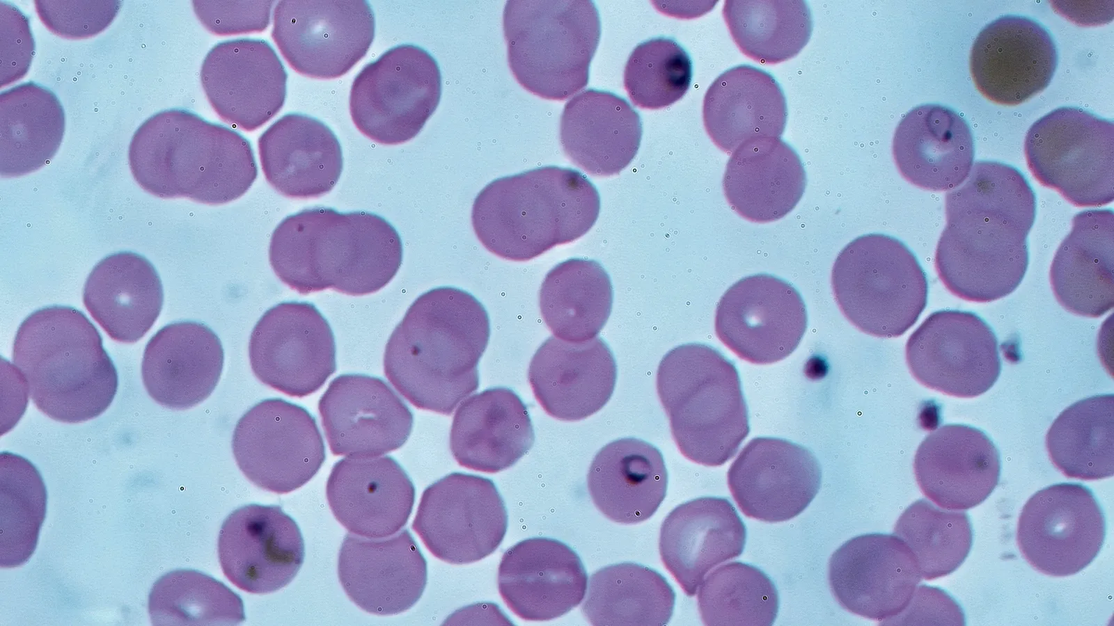
In practice the colours do the work. Giemsa is a nucleic acid stain, and in a blood film red cells come out pink while the chromatin of white-cell nuclei turns magenta (Giemsa stain overview). A malaria parasite inside a red cell has its own nucleus, so its chromatin takes up the stain too and shows as a small dark purple dot against the pink cell.
That purple is called the Romanowsky effect: shades of purple in chromatin that neither dye gives alone, now attributed to azure B and eosin acting together (Romanowsky stain overview).
Is Giemsa stain still used today?
Yes. The US Centers for Disease Control and Prevention says microscopy remains the "gold standard" for laboratory confirmation of malaria, using blood smears stained with Giemsa or Wright-Giemsa and examined with a 100X oil immersion objective. Two smears are made from the same blood (CDC DPDx malaria page):
a thick smear, to detect whether parasites are present at all;
a thin smear, to identify the species and the share of red cells infected.
CDC lists four true human malaria parasites, *P. falciparum*, *P. vivax*, *P. ovale* and *P. malariae*, plus *P. knowlesi*, a monkey parasite sometimes found in people. CDC's own blood staining procedure dilutes stock Giemsa to 2.5% in a buffer at pH 7.2 and leaves slides in it for 45-60 minutes.
The stain also travelled beyond malaria. Barcia notes it became the basic stain of the Kiel classification of lymphomas, and Giemsa banding (G-banding) of chromosomes is used to build karyograms (Giemsa stain overview).
How do you read a Giemsa-stained smear?
If a school lab or a Class 11-12 practical puts a stained blood slide under your microscope, work through it in this order. Our guide to stains used in biology practicals covers the other common ones.
Start at low power and find the thin, feathered edge of the smear, where cells lie in a single layer.
Check the background colours: red cells pink, white-cell nuclei magenta to purple. If the whole slide looks one colour, the staining went wrong, which is the exact problem Giemsa set out to fix.
Move to high power and look inside red cells. Specks of stain or dust can mimic parasites, so focus up and down to check what lies inside the cell.
Look for a ring: a purple chromatin dot with a thin loop of blue around it.
Write down what you see before you name it. "Purple dot with a blue ring inside a red cell" is an observation; "*P. falciparum*" is a conclusion only a trained microscopist should make.
A slide in a school lab is for learning. A fever after a trip, or during the monsoon, needs a doctor and a proper test, not a guess.
If the chemistry behind the colours is the shaky part, the Fermi Concept Library covers every concept in physics, chemistry, biology and mathematics, so you can look up nucleic acids or buffers on their own.
Next, follow the parasite you just spotted through its full journey in our malaria Plasmodium life cycle explainer.