
On this page
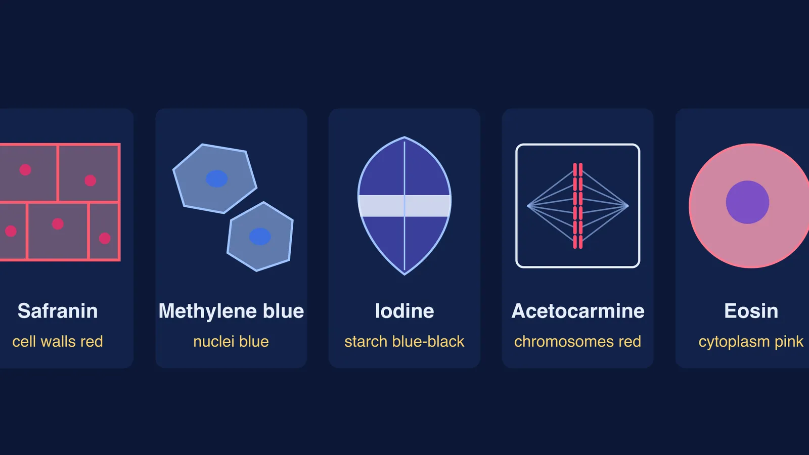
Your examiner slides a stained mount under the microscope and asks one question: which stain, and why? This guide covers five stains that come up in school biology practicals: safranin, methylene blue, iodine, acetocarmine and eosin. Each one colours something different, and that colour is a clue to what you are looking at.
Which stain is used for onion peel and cheek cell slides?
In the NCERT Class 9 science lab manual, Experiment 18 (a stained temporary mount of onion peel) lists "methylene blue (or safranin)" as the stain. The peel sits in dilute stain for about 3 minutes, is rinsed in water and is mounted in glycerine.
Safranin stains cell walls that contain lignin red, and it can also stain walls that contain cutin, because it is not specific to one chemical, according to plant morphology course notes from UKF Nitra. That is why plant cell walls, and lignified xylem in stem or root sections, stand out.
Experiment 19 in the same Class 9 lab manual is the cheek cell mount: gently scrape the inside of your cheek with a clean toothpick, spread the material in a drop of water on a slide, add a drop of methylene blue, and after about 3 minutes add glycerine and a cover slip.
Methylene blue gives a blue colour to both nuclei and cytoplasm and makes the nuclei more visible, as the methylene blue entry on Wikipedia summarises. The manual's note for teachers adds that organelles are not visible in this preparation because they are too small and are not stained by methylene blue.
Which stain shows starch in a leaf?
In Experiment 24 of the NCERT Class 10 science lab manual, part of a leaf on a de-starched plant is covered with black paper and kept in bright sunlight for about two hours. The leaf is softened in boiling water, decolourised in alcohol warmed in a water bath, then dipped in iodine solution for about five minutes. The manual states that a blue-black colour confirms starch: the exposed part turns blue, the covered part does not.
Which stain is used for the onion root tip mitosis slide?
Acetocarmine is a 1% solution of carmine in 45% acetic acid used to stain plant chromosomes, according to the Wheat Genetics Resource Center at Kansas State University. Carmine itself is a bright-red pigment (Wikipedia: Carmine).
The Virtual Labs mitosis experiment explains why the onion root tip is used: it grows rapidly, so many cells are caught in different stages of mitosis. Its procedure adds 2-3 drops of acetocarmine to the root tip on a slide and warms it gently for about one minute.
What does eosin stain on a permanent slide?
None of the NCERT experiments above uses eosin. Eosin is an acidic, negatively charged dye that stains basic structures red or pink, explains the University of Leeds histology guide. Paired with haematoxylin, which stains acidic structures such as nuclei purplish blue, it makes H&E, which the same guide calls the most commonly used staining system. Pink cytoplasm with purple-blue nuclei on a permanent slide? Think H&E.
Why does each stain colour a different part?
A stain binds some chemicals more than others. Per the Leeds guide, eosin stains most cytoplasmic proteins pink because they are basic, while haematoxylin, a basic dye, stains the DNA-rich nucleus. Carmine is also a basic dye (Kansas State), which fits its use on chromosomes. Safranin follows lignin and cutin, and iodine reacts with starch.
One line to remember: red walls, blue cheeks, black starch, red chromosomes, pink cytoplasm.
How do you answer "which stain and why" in a viva?
Name the stain, say what it colours, and link that to the aim. Three worked answers:
Onion peel: "I used safranin. It stains the cell walls red, so I can see that the cells are rectangular, compactly arranged and have no intercellular spaces."
Cheek cells: "I used methylene blue. It colours the nucleus and cytoplasm blue. There is no cell wall, so the polygonal outline is the cell membrane."
Root tip: "I used acetocarmine. It stains the chromosomes red, and the root tip divides rapidly, so I can find cells in different stages of mitosis."
Answers 1 and 2 use the results in the Class 9 lab manual. Its precautions and steps for these two slides include:
Rinse off excess stain in water.
Avoid over-staining or under-staining.
Avoid air bubbles when you lower the cover slip.
Blot excess glycerine from the edges with blotting paper.
For the full method behind each slide, see our guides to preparing onion peel and cheek cell slides, the onion root tip mitosis practical and the leaf starch test practical.
If acidic and basic dyes or the stages of mitosis still feel fuzzy, the Fermi Concept Library has a lesson for every biology concept, and Fermi's AI tutor checks your understanding as it goes, much as a viva does.
Before your practical, write the five stains on one card and say each answer out loud once.