
On this page
Before 1927, a plain X-ray of the head showed the skull and little else, because brain, blood and vessel walls blur into one grey shadow. Egas Moniz, a Portuguese neurologist (1874 to 1955), wanted to use X-rays to make the brain's vessels visible and so locate brain tumours. His idea was to fill the arteries with something X-rays cannot easily pass through, then look for places where a tumour had pushed them aside.
What did Egas Moniz do in 1927?
A 2017 review in Frontiers in Neuroanatomy describes how Moniz tested three possible dyes, lithium bromide, strontium bromide and sodium iodide, in cadavers, animals (rabbits and dogs) and patients. Lithium bromide was irritant and caused brief pain. Strontium bromide caused a feeling of warmth when injected quickly. A 25% solution of sodium iodide was painless and gave optimal contrast.
The first human trials went badly: in his first four patients the images failed, and in a second series one patient died of a stroke 8 hours after the procedure. Moniz blamed very thin needles that may have slipped out of the artery, so he switched to surgically exposing the internal carotid artery in the neck.
In June 1927 it worked. The review describes a 20-year-old man with a pituitary tumour whose brain arteries showed clearly on the film. Moniz presented the result to the Neurological Society in Paris on 7 July 1927. The review and a 2024 review in Cureus both describe the clinical logic: a tumour displaces nearby vessels, so a shifted artery on the film tells doctors where the tumour sits.
His carotid injections filled the ring of arteries described in our post on Thomas Willis and the circle of Willis.
Why does iodine show up on an X-ray?
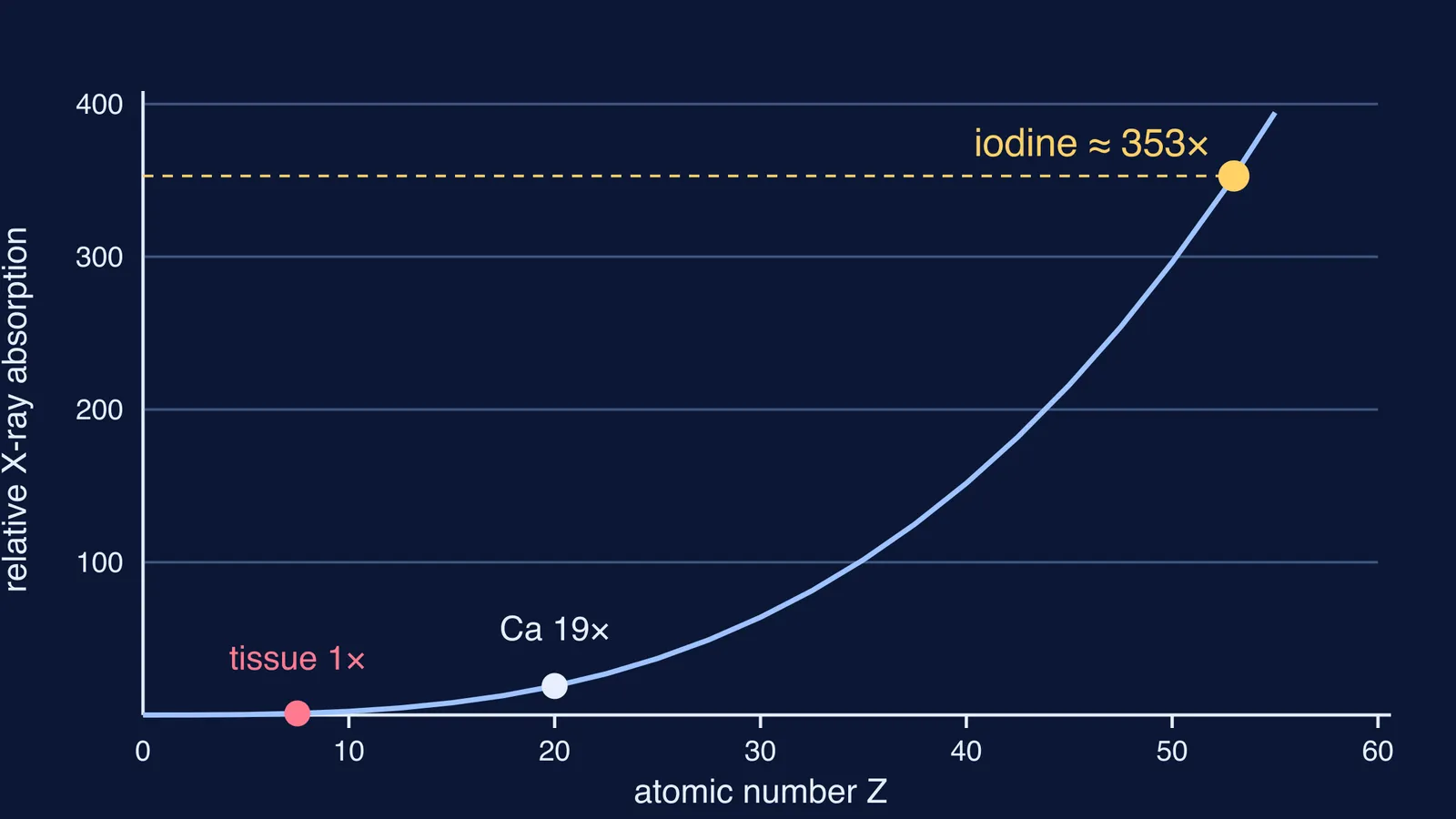
Iodine has atomic number 53, much higher than most tissues in the body, so it absorbs X-rays far more strongly, as Radiopaedia's entry on iodinated contrast media explains. Its K-edge, the energy needed to remove an electron from its innermost shell, is 33.2 keV, similar to the average energy of diagnostic X-rays, which makes absorption even more likely.
At these energies the main absorption process is the photoelectric effect, whose probability rises roughly as Z³ and falls roughly as 1/E³, according to Seibert and Boone in the Journal of Nuclear Medicine Technology. You can work out the size of the effect yourself:
Soft tissue has an effective atomic number of about 7.5; iodine has 53.
Divide and cube: (53 ÷ 7.5)³ ≈ 353. Seibert and Boone round this to about 350 times more absorption for the same thickness at the same energy.
Try calcium (Z = 20), which helps make bone visible: (20 ÷ 7.5)³ ≈ 19.
Now change the energy instead: doubling E divides the photoelectric term by 2³ = 8.
The same reasoning explains how X-rays show bones but not the soft organs around them.
If the photoelectric effect still feels hazy, the Fermi Concept Library gives every concept in physics, chemistry and biology its own lesson, so you can revisit the idea before trying the Z³ sums again.
Is cerebral angiography still used today?
Yes. The Frontiers review concludes that for specific and frequent conditions, brain angiography "still has a diagnostic and therapeutic application, which remains unsurpassed". In the modern version described by RadiologyInfo, a thin plastic catheter is inserted into an artery in the leg or arm, and contrast material is injected through it. Doctors use it to look for aneurysms, arteriovenous malformations, narrowed arteries, clots and strokes. RadiologyInfo notes that stroke is a possible but uncommon complication.
For the maths behind another way of seeing inside the head, read about Allan Cormack and the mathematics of CT.
Why is Moniz's Nobel Prize controversial?
The Frontiers review records that Moniz was nominated for the Nobel Prize for angiography in 1928, 1932 and 1937 without winning. He won in 1949, shared with Walter Rudolf Hess, for prefrontal leucotomy, an operation that cut connections between the prefrontal cortex and other parts of the brain. The Cureus review notes that he was criticised for downplaying complications, inadequate documentation and poor patient follow-up, and that the arrival of chlorpromazine in 1952 led to the operation's decline.
How can you use Moniz's method to think through a problem?
Five questions work for any "how could we see X?" problem:
What do you want to see, and why is it invisible now? (Vessels and brain absorb X-rays alike.)
Which physical property creates contrast? (Photoelectric absorption, rising roughly as Z³.)
Which material has lots of that property? (Iodine, Z = 53.)
Is it safe enough? (Lithium bromide hurt; sodium iodide did not.)
How will you know it worked? (Vessels appear on the film, and shifted vessels point to a tumour.)
Next time a textbook says something "shows up" on a scan, ask which of the five steps made that possible.