On this page
If you have ever broken an arm or visited a dentist, you have seen the stark black-and-white image of your own skeleton. While it looks like a photograph, an X-ray is actually a map of shadows. It records what the radiation failed to pass through.
The physics behind this everyday medical tool connects the electromagnetic spectrum to the atomic structure of the human body.
How does an X-ray show bones inside the body?
An X-ray machine does not take a picture in the way a camera does. Instead, it fires a beam of high-energy radiation through the body toward a detector (traditionally photographic film, but now usually a digital sensor) on the other side.
Because the human body is not uniform, different tissues absorb different amounts of this radiation. X-rays are a form of high-energy electromagnetic radiation with wavelengths ranging from roughly 10 nanometers to 10 picometers. This short wavelength gives them the power to penetrate living tissue.
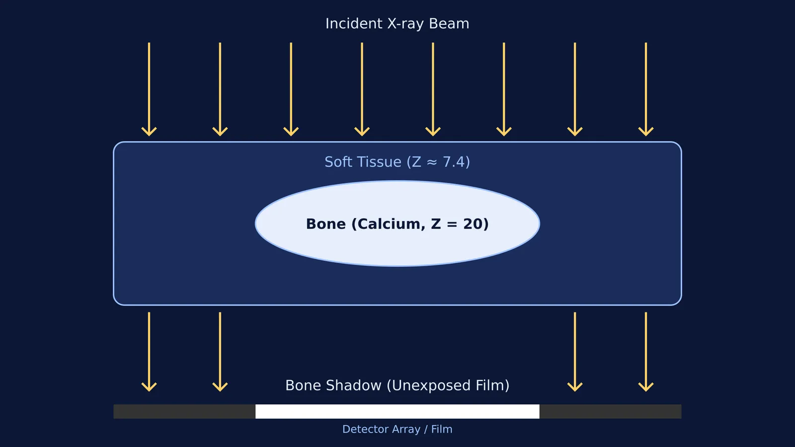
When the beam passes through the body, your skin, muscles, and organs let most of the radiation pass through. This radiation hits the detector, turning it black. Your bones, however, block the radiation. The X-rays never reach the detector behind the bone, leaving those areas unexposed and white. You are essentially looking at a shadow cast by your skeleton.
Why does bone absorb more X-rays than soft tissue?
The stark contrast between bone and soft tissue comes down to atomic numbers.
At the photon energies typically used for medical imaging (ranging from 20 keV to 150 keV), X-rays interact with heavier elements primarily through the photoelectric effect. In this process, an X-ray photon transfers all its energy to an inner-shell electron of an atom, ejecting the electron and being completely absorbed.
The probability of this photoelectric absorption happening is highly sensitive to the atomic number (Z) of the target material. Specifically, the absorption cross-section is proportional to the cube of the atomic number (Z³).
Soft tissue is mostly composed of water and organic molecules made of hydrogen (Z=1), carbon (Z=6), and oxygen (Z=8). Its overall effective atomic number is approximately 7.4.
Bone is a dense matrix containing a high concentration of calcium, which has an atomic number of 20.
Because 20³ (8,000) is substantially larger than 7.4³ (about 405), bone is vastly more efficient at absorbing X-rays than the surrounding tissue. This steep mathematical difference is why the ribs and spine stand out so clearly against the lungs and heart.
When physics meets study habits
Understanding how X-rays interact with matter is a staple of modern physics units. It ties together the electromagnetic spectrum, atomic models, and X-rays and Moseley's law.
However, reading about the photoelectric effect is one thing; picturing how it builds an image is another. If you are preparing for exams and find these atomic interactions hard to visualise, the Fermi Simulation Lab offers interactive simulations for the ideas that are easier to see than to read. You can adjust the density and atomic number of target materials to watch how the absorption changes in real time.
A practical framework: How to look at an X-ray
While diagnosing an injury is strictly a doctor's job, understanding the basic structure of a radiograph helps demystify the process. Medical students are often taught the "ABC" framework to systematically read a bone X-ray:
Alignment: Do the bones line up naturally? A sudden shift in the normal axis can indicate a fracture or dislocation.
Bones: Trace the outline (cortex) of the bone. The edges should be smooth and continuous. A jagged break or a "step" in the bright white outline is the most common sign of a fracture.
Cartilage and joint spaces: Cartilage has a low atomic number, similar to water, so it does not show up as white on an X-ray. Instead, doctors look at the empty black space between bones. If the gap is too narrow, the invisible cartilage may be worn away.
By applying the physics of atomic absorption to the anatomy of the human body, an X-ray turns a simple shadow into one of the most powerful diagnostic tools in medicine.