
On this page
When a dicot stem or root thickens, its single layer of epidermis cannot keep up. The plant replaces it with a new protective tissue made by a second lateral meristem, the cork cambium. If lateral meristems are new to you, start with our guide to apical, intercalary and lateral meristems.
What is the periderm, and what are its three layers?
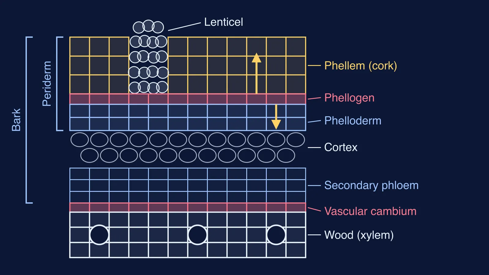
The periderm is the protective tissue that forms as stems and roots grow in girth and takes over from the epidermis and endodermis, according to a 2022 review in the Annual Review of Plant Biology by Olga Serra, Ari Pekka Mähönen, Alexander Hetherington and Laura Ragni. The same review notes that a wound periderm also forms to heal and protect injured tissue. Its three layers are:
Phellogen (cork cambium): the meristem, a thin layer of dividing cells that makes the other two.
Phellem (cork): the cells the phellogen makes towards the outside, dead at maturity with suberised walls (Peter v. Sengbusch, Botany online).
Phelloderm: the cells made towards the inside, living parenchyma usually 1 to 4 cell layers thick, if present at all (plant anatomy lecture notes, Nickrent lab, Southern Illinois University).
A memory hook: phellogen generates; phelloderm sounds like dermis, the layer under your skin, so it sits inside.
Where does cork cambium come from in secondary growth?
The vascular cambium has a head start: in a dicot stem it already sits between xylem and phloem in each open vascular bundle, as NCERT Class 11 Biology, Chapter 6 describes. The cork cambium does not. It forms later, during thickening, from living cells that start dividing again. Where depends on the organ (SIU lecture notes):
In stems: from the epidermis, or from parenchyma or collenchyma just beneath it.
In roots: in the pericycle, or near the surface.
Once it exists, the phellogen adds phellem on the outside and phelloderm on the inside. Phellem cells are generated outwards, are non-living and have suberised walls (Botany online), so tissues left outside a periderm are isolated from the living stem. The SIU notes add that a new periderm can form each year, in successively deeper layers beneath the first. For the vascular cambium side of the story, see secondary growth in dicot stems, step by step.
How are bark and lenticels related to the periderm?
Bark is all the tissue outside the vascular cambium, with an inner living part and an outer dead part (SIU lecture notes). So bark is a region, not one tissue: it includes the secondary phloem, any primary tissue still there (such as cortex), and the periderm. Its dead outer part, the rhytidome, is made of layers isolated by periderms plus periderm that has stopped growing.
Lenticels are openings in the periderm of stems and roots used for gas exchange, according to the same notes. They first appear under stomata, where the phellogen is more active and makes loose filling (complementary) cells instead of tightly packed cork. Botany online describes them as letting gases pass between the metabolically active cells below the bark and the air. To compare them with stomata, read how lenticels and stomata manage gas exchange.
How do you draw and label the periderm?
Try this worked example: one strip of an older dicot stem, outside at the top, like the diagram above this post.
Draw a thin row of small cells and label it phellogen. In a stem, place it just under where the epidermis was.
Above it, draw 3 or 4 rows of tightly packed, empty, box-shaped cells. Label them phellem (cork).
Below it, draw 1 or 2 rows of cells with nuclei. Label them phelloderm.
Bracket those three layers as periderm.
Below, add cortex, secondary phloem, the vascular cambium and wood. Bracket everything above the vascular cambium as bark.
Break the cork in one place, fill the gap with loose round cells bulging outward, and label it lenticel.
Then test yourself without looking:
Which layer is alive, phellem or phelloderm? Phelloderm.
Which way does the phellogen make cork? Outwards.
Where does cork cambium form in a root? In the pericycle, or near the surface.
Is secondary phloem part of the bark? Yes: it lies outside the vascular cambium.
If a question trips you up, the Fermi Concept Library covers every concept in biology, each with its own lesson and its place in the map, so you can see how periderm connects to meristems and vascular tissue. Fermi's AI tutor draws the figure as it explains and checks your understanding as it goes.
Is cork cambium still in NCERT Class 11 Biology?
The 2026-27 reprint of NCERT Class 11 Biology, Chapter 6 has no section on secondary growth and does not mention cork cambium or periderm; its summary notes that secondary growth occurs in most dicotyledonous roots and stems. Your board or entrance exam may still expect more, so check the syllabus your exam body publishes for your year before you drop it.
Next step: draw the strip from memory once, then check it against the six steps above.