
On this page
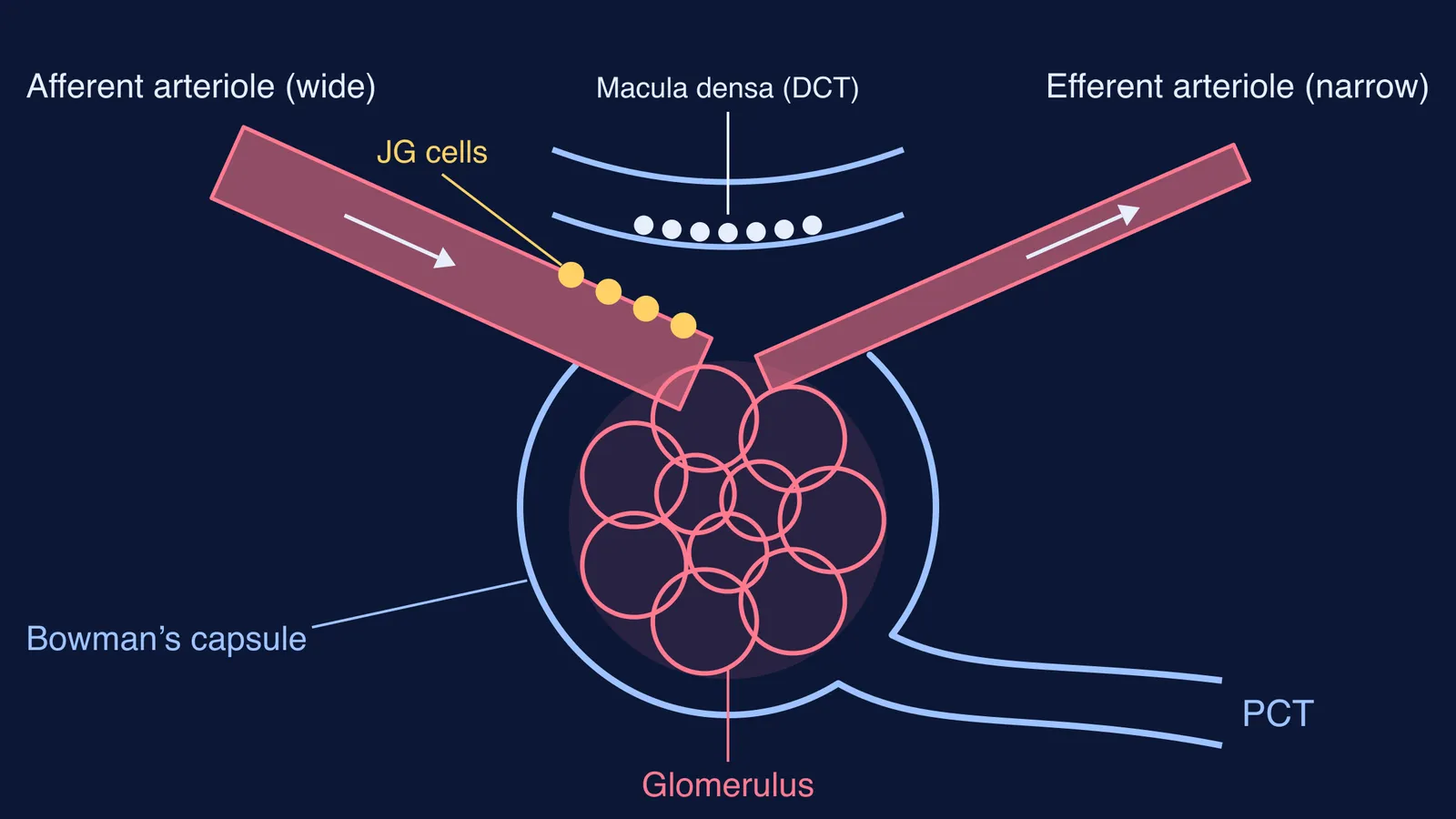
Most capillary beds sit between an arteriole and a venule. The glomerulus is different: it sits between two arterioles, the afferent one bringing blood in and the efferent one taking it out (StatPearls: Histology, Kidney and Glomerulus). NCERT's Class 11 chapter describes the glomerulus as a tuft of capillaries formed by the afferent arteriole, a fine branch of the renal artery, with blood carried away by an efferent arteriole (NCERT Biology Class 11, Chapter 16). Their difference in width helps keep the pressure inside the glomerulus high enough to filter.
Why is the afferent arteriole wider than the efferent arteriole?
Think of a garden hose with your thumb over the end: the exit is squeezed, so pressure inside rises. In the glomerulus, a wide inlet lets blood in easily, a narrower outlet resists its leaving, and pressure in the capillaries between them stays high.
Glomerular capillary hydrostatic pressure is the principal filtration force and measures about 55 mm Hg (StatPearls: Physiology, Renal). Two forces push back: the fluid pressure inside Bowman's capsule and the colloid osmotic pressure of plasma proteins in the capillary. The balance of all of them is the net filtration pressure.
The glomerular filtration rate (GFR) in a healthy person is about 125 ml per minute, or 180 litres a day (NCERT Class 11, Chapter 16). Our explainer on GFR and the ultrafiltration membrane covers the three layers that pressure pushes fluid through.
A footnote: a 2024 review in Frontiers in Physiology, citing earlier measurements, reports that efferent arterioles are narrower than afferent ones in the outer and middle cortex, but about the same width in the juxtamedullary cortex, close to the medulla (Kuang, Ahmetaj and Qu, 2024). For school exams, the textbook answer stands.
How does a narrow efferent arteriole raise glomerular pressure?
The same review cites Poiseuille's equation: the resistance of a tube is inversely proportional to the fourth power of its radius (Kuang, Ahmetaj and Qu, 2024). A simplified worked example:
Take two arterioles of equal length, and give the efferent half the radius of the afferent.
Resistance scales as 1/r⁴, so the efferent has 2⁴ = 16 times the resistance of the afferent.
The glomerulus sits between the two, like the junction of two resistors in series. Most of the pressure drop happens across the larger resistance, which is now downstream.
Pressure at the junction = outflow pressure + 16/17 of the total drop, not half of it. The glomerulus keeps most of the pressure the afferent delivers.
Real arterioles differ in length and fluid leaves along the way, so treat this as a model, not a measurement.
During efferent constriction GFR rises while renal plasma flow falls, and angiotensin II preferentially constricts efferent arterioles to keep filtration going when renal blood flow is low (StatPearls: Renal Blood Flow and Filtration). In the other direction, macula densa signals contract smooth muscle in the afferent arteriole to bring a high GFR back to normal (StatPearls: Physiology, Glomerular Filtration Rate).
Where is the juxtaglomerular apparatus located?
NCERT defines the juxtaglomerular apparatus (JGA) as a special sensitive region formed by cellular modifications in the distal convoluted tubule and the afferent arteriole at the location of their contact (NCERT Class 11, Chapter 16). In practice it sits at the vascular pole, in the angle where the two arterioles meet the capsule.
JG (granular) cells: in the wall of the afferent arteriole; they release renin when stretch falls (StatPearls: GFR).
Macula densa: tubule cells pressed against the vascular pole that sense salt delivery. StatPearls places them in the thick ascending limb; NCERT says distal convoluted tubule, so write it NCERT's way in board exam answers.
NCERT's one-line function: a fall in GFR can activate the JG cells to release renin, which can stimulate glomerular blood flow and bring GFR back to normal. What renin sets off next is in our post on ADH, the RAAS and ANF.
How do you draw the labelled sketch?
Redraw the diagram above from memory, then check.
Draw Bowman's capsule as a cup, open at the top (vascular pole), with the PCT leaving the bottom.
Fill it with a knot of capillary loops: the glomerulus.
Add a thick tube entering one side of the pole: afferent arteriole, arrow pointing in.
Add a tube half as wide leaving the other side: efferent arteriole, arrow pointing out.
Draw a short piece of distal tubule in the angle between them. Shade the cells touching the pole: macula densa.
Dot the afferent wall next to the pole: JG cells. Bracket them with the macula densa as JGA.
For the whole nephron, follow our nephron sketch with Bowman's capsule and tubule.
If the pressure idea still feels slippery, the Fermi Concept Library gives every concept in physics, chemistry, biology, mathematics and computing its own lesson, so you can trace resistance into filtration. To test yourself, the Fermi Problem Library has worked problems with a step-by-step solver that explains the reasoning rather than printing an answer.