On this page
Paul Langerhans was born in Berlin in 1847. His father was a physician and a friend of the pathologist Rudolf Virchow, according to a 2025 history of Langerhans in the journal Pathologica. He studied first in Jena under the zoologist Ernst Haeckel, then in Berlin with Virchow.
He made his first discovery before he graduated. At 21 he used a gold chloride stain to find branching cells in the outer layer of the skin, now called Langerhans cells (Pathologica). He thought they were nerve receptors. It took more than a century to work out their real job in the immune system.
What did Langerhans discover in the pancreas in 1869?
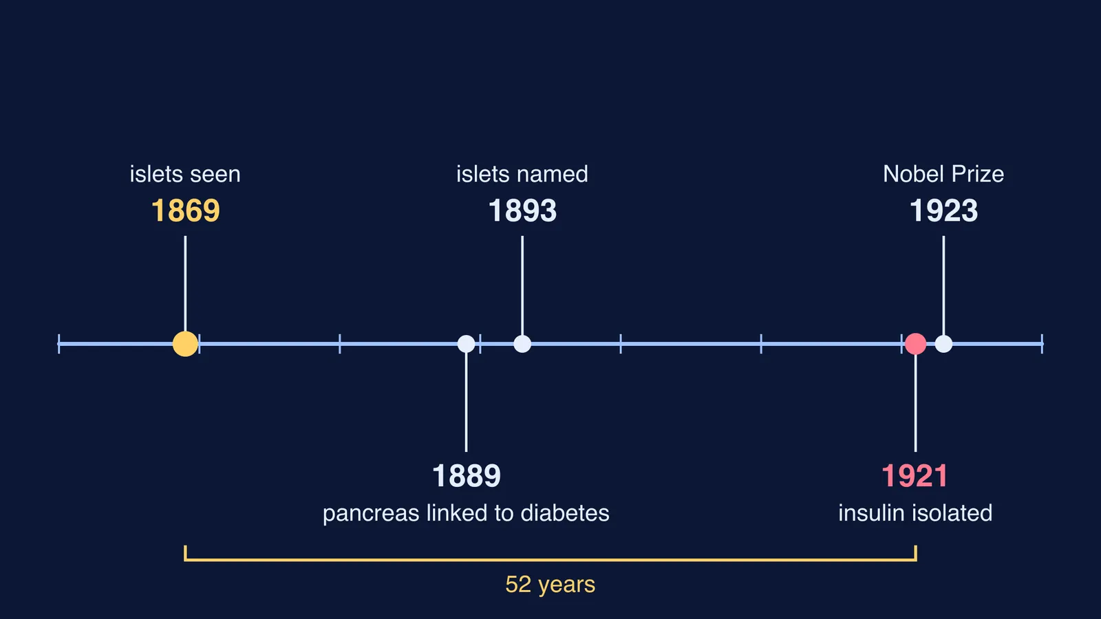
In 1869, aged 22, Langerhans defended his doctoral thesis, *Beiträge zur mikroskopischen Anatomie der Bauchspeicheldrüse* (Contributions to the microscopic anatomy of the pancreas), under Virchow's supervision (Pathologica, 2025). At the time the pancreas was still widely thought of as a kind of salivary gland in the abdomen.
His tools were basic. He worked on rabbit pancreas with no microtome to cut thin sections and no haematoxylin-eosin stain. Instead he hardened the gland in Müller's fluid, carefully macerated it, and stained it with gold chloride, carmine and osmic acid (Pathologica).
Among the tissue that makes digestive enzymes, he noticed small clusters of different cells spread through the whole gland. They had no connection to the ducts. In the English translation of his thesis, quoted in Pathologica, he described them as "small irregular polygonal structures with perfectly brilliant cytoplasm free of granules, distinct round nuclei of moderate size". He did not draw them, did not name them, and did not say what they did.
How did the islets of Langerhans get their name?
The French histologist Édouard Laguesse found the clusters again in an adult human pancreas, called them "îlots de Langerhans" in 1893, and proposed that they were the source of an internal secretion (Pathologica).
A history in Frontiers in Endocrinology sets out the steps that followed:
1889: Joseph von Mering and Oskar Minkowski removed a dog's pancreas, linking the organ to diabetes (we tell that story in how Minkowski and von Mering linked the pancreas to diabetes).
1909: Jean de Meyer proposed the name "insulin" for the islets' still-unseen secretion (some histories credit Edward Sharpey-Schafer instead).
1921: Frederick Banting and Charles Best tied off the pancreatic ducts so the enzyme tissue wasted away, then extracted insulin.
January 1922: Leonard Thompson, a 14-year-old at Toronto General Hospital, became the first patient treated.
1923: Banting and John Macleod received the Nobel Prize in Physiology or Medicine.
Today we know the islets take up only 1-2% of the volume of the human pancreas, according to BioNumbers, citing a histology textbook. Inside them, A cells secrete glucagon, B cells insulin and D cells somatostatin, as a review in the Journal of Histochemistry and Cytochemistry summarises. For the exam version of how alpha and beta cells balance blood sugar, see our guide to alpha and beta cells in the islets of Langerhans.
Langerhans did not see any of this. He became a full professor at 27, and weeks later learned he had tuberculosis (Pathologica). He moved south for his health, kept studying marine animals in Madeira, and died in Funchal on 20 July 1888, aged 41.
What can a student learn from Langerhans's thesis?
His thesis is useful because he kept two things apart: what he saw and what he could explain. Here is a four-step method based on what he did.
Scan the whole field first. He noticed the clusters because he looked across the gland, not at one spot.
Describe before you name. Shape, size, colour, texture, position. His words were plain: polygonal, granule-free, round nuclei.
Compare with the neighbours. What makes this patch different from the tissue around it? For him, it was the missing link to the ducts.
Write the open question down. "I cannot yet explain this" is a valid line. It tells you, and your examiner, exactly what to look up next.
Here is a worked example for a pancreas slide in your biology practical record.
What I see: pale, rounded patches of cells among darker-staining clusters of enzyme-making cells.
How it differs: no duct opens into the pale patches.
My question: if these cells have no duct, where does their product go?
What I then checked: it goes straight into the blood, which is what makes the islets endocrine tissue.
If step four keeps sending you back to gaps about hormones and glands, the Fermi Concept Library has every concept in biology, each with its own lesson and its place in the map. Feedback between insulin and glucagon is easier to see than to read, which is what the interactive simulations in the Fermi Simulation Lab are for.
Next time a slide shows you something you cannot explain, write it down the way a 22-year-old did in 1869.