

On this page
In the spring of 1889 at the University of Strasbourg, physician Oskar Minkowski and pharmacologist Joseph von Mering debated whether animals could absorb dietary fat without a pancreas. Von Mering believed surgical removal of the entire organ was fatal. Minkowski, confident in his operative skill, removed the pancreas from a dog that evening in Bernhard Naunyn’s clinic. The animal survived, but soon developed extreme thirst, ravenous hunger, and copious sweet urine. That unexpected finding proved that the pancreas does far more than produce digestive enzymes: it releases a vital blood-borne agent that regulates blood sugar.
Why did Minkowski and von Mering remove a dog's pancreas?
In the late nineteenth century, physiologists viewed the pancreas strictly as an exocrine digestive gland. Claude Bernard had demonstrated that pancreatic juice digests fats, starches, and proteins in the gut. Joseph von Mering wanted to test whether an animal deprived of pancreatic secretions could still absorb dietary lipids.
Earlier surgical attempts had failed because experimental animals quickly died of infection or shock. Von Mering considered total pancreatectomy impossible, but Minkowski had successfully performed delicate organ surgeries in birds. In Naunyn’s laboratory, Minkowski cleanly extirpated the canine pancreas while preserving the duodenum and nearby blood vessels, creating a living subject to study fat absorption.
How did Minkowski discover sugar in the dog's urine?
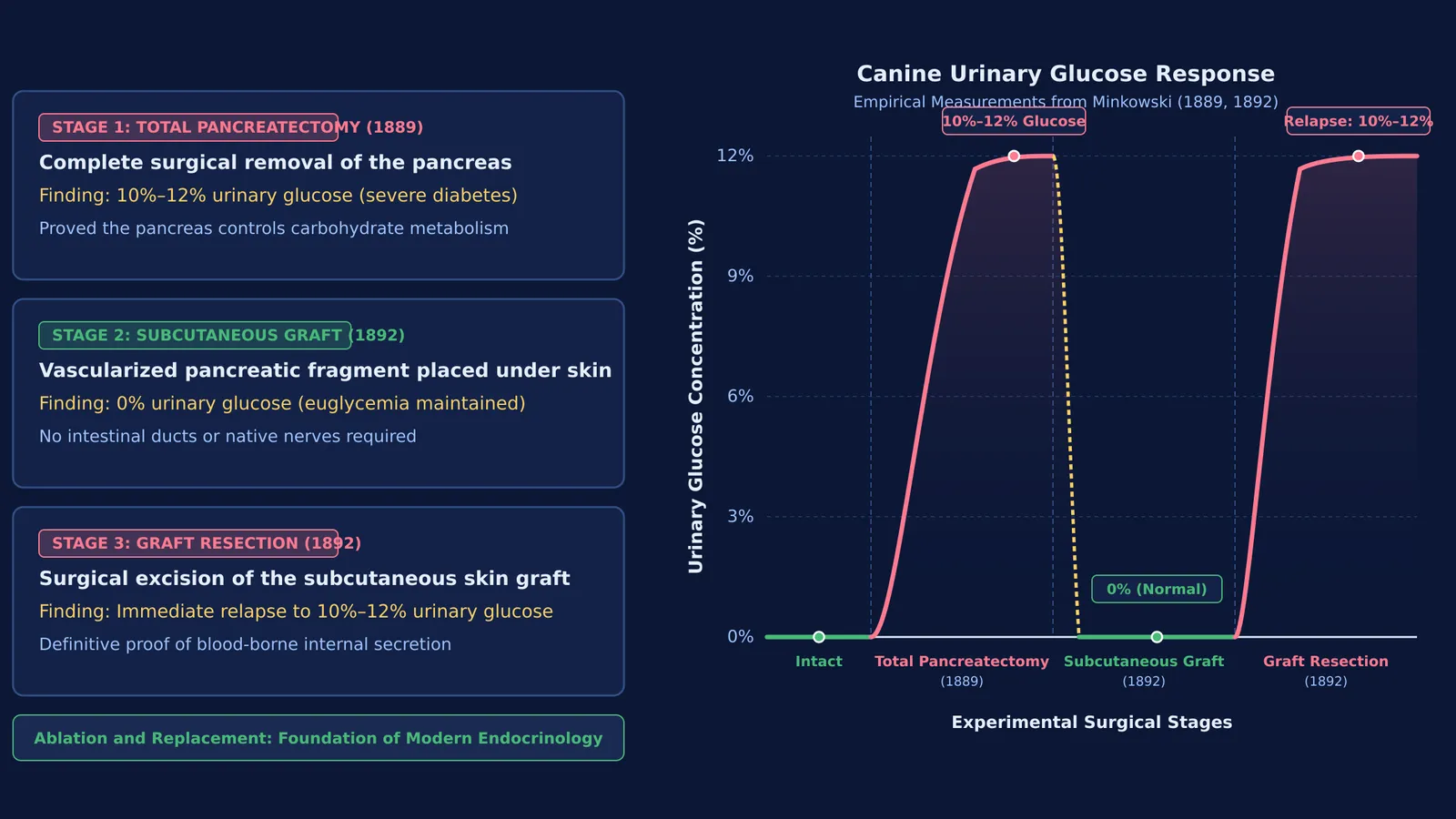
The discovery of diabetes was completely accidental. Following surgery, the house-trained dog began urinating copiously across the floor. Minkowski collected the urine and conducted Trommer’s and Fehling’s reduction tests, verified by polarimetry. The urine contained a massive concentration between 10% and 12% glucose, as published in their 1890 report in the Archiv für experimentelle Pathologie und Pharmakologie.
The animal displayed severe diabetes mellitus: polyuria, constant thirst, voracious hunger, and rapid wasting. Because Minkowski worked in the clinic of Bernhard Naunyn—a leading authority on clinical diabetes—he recognized the signs immediately. Until then, medicine blamed diabetes on disorders of the liver, kidneys, or nervous system. Minkowski and von Mering showed that losing the pancreas alone caused the disease.
How did the 1892 pancreatic graft prove internal secretion?
Critics argued that diabetes arose from damage to abdominal nerve plexuses or the stoppage of digestive secretions. In his 1893 treatise in the Archiv für experimentelle Pathologie und Pharmakologie, Minkowski documented that fasting depancreatized dogs maintained a dextrose-to-nitrogen ratio of 2.8:1, showing the body degraded structural proteins into sugar. To definitively prove an internal secretion, he designed a two-stage graft experiment in 1892.
Minkowski removed the abdominal pancreas but grafted a vascularized piece of pancreatic tissue under the dog’s skin. The subcutaneous graft lacked ducts to the intestine and had no abdominal nerves. Yet as long as this graft lived, the dog remained entirely healthy. When Minkowski later excised the skin graft, severe glycosuria returned within hours. This proved that the pancreas controls glucose via an internal secretion released into the blood.
What does Minkowski's work mean for modern endocrine biology?
Minkowski’s work established the core logic of endocrinology: remove a gland to demonstrate deficiency, then replace it to restore normal metabolism. His finding guided subsequent research, culminating in Frederick Banting and Charles Best's isolation of insulin in 1921. Historical reviews in the Journal of Community Hospital Internal Medicine Perspectives and Acta Diabetologica recognize Minkowski's depancreatized dog as the foundation of modern diabetes therapy.
The distinction between duct-borne enzymes and blood-borne messengers also inspired William Bayliss and Ernest Starling's discovery of secretin, which coined the term hormone. Today, biotechnology manufactures human insulin through recombinant insulin production techniques.
When you study glandular physiology and feedback loops, the Fermi Concept Library covers every concept in physics, chemistry, biology, mathematics and computing, each with its own lesson and its place in the map. For mechanisms that are hard to picture from static diagrams, the Fermi Simulation Lab offers interactive simulations for the ideas that are easier to see than to read.
How do researchers test endocrine gland function?
Minkowski's experiments, synthesized in his 1893 Archiv für experimentelle Pathologie und Pharmakologie monograph, established the classical ablation-and-replacement framework for endocrine physiology:
Surgical ablation: Completely remove the candidate organ to observe which systemic functions fail.
Clinical phenotyping: Measure key biomarkers, such as urinary glucose and fluid output, to identify specific hormone loss.
Ectopic transplantation: Graft the tissue to a distant site without ducts or native nerves, testing whether blood circulation alone restores function.
Secondary resection: Remove the ectopic graft to verify that deficiency symptoms return, confirming tissue-dependent control.


