On this page
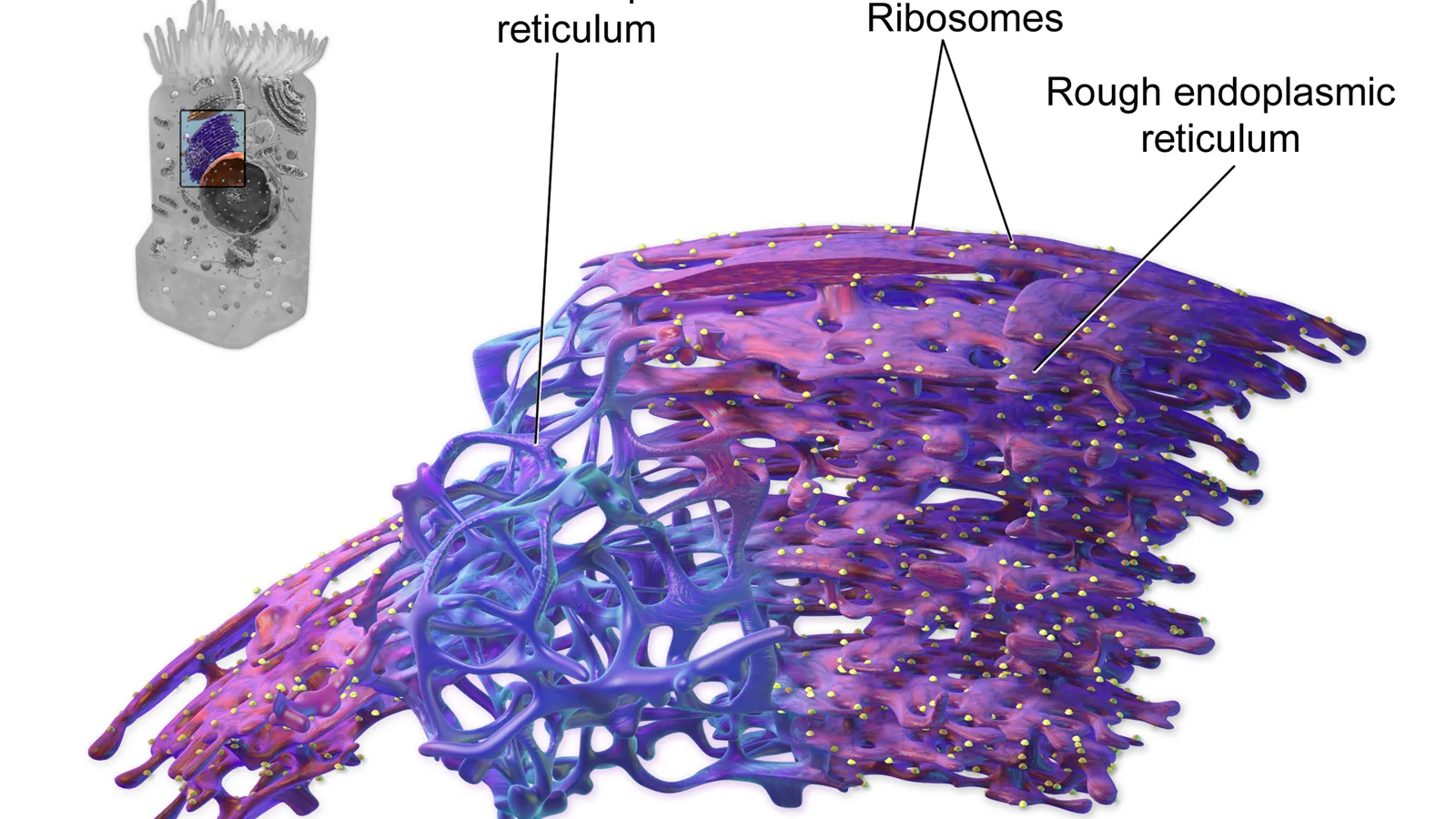
Every Class 11 student draws the endoplasmic reticulum (ER) as a maze of folded membranes next to the nucleus. The NCERT chapter Cell: The Unit of Life says it plainly: electron microscope studies of eukaryotic cells reveal a network of tiny tubular structures in the cytoplasm, called the endoplasmic reticulum. It does not say who first described and named that network. That was Keith Porter, born in Yarmouth, Nova Scotia, in 1912, according to Encyclopedia.com's entry on Porter.
Who took the first electron micrograph of a whole cell?
In 1945, Keith Porter, Albert Claude and Ernest Fullam published "A study of tissue culture cells by electron microscopy" in the Journal of Experimental Medicine. Their abstract is short: cells from chick embryos were grown by a tissue culture technique in a state suitable for electron microscopy, and the micrographs showed details of cell structure not revealed by other methods.
Porter grew embryonic chick cells on a thin plastic (polyvinyl) film, peeled them off, put them on a wire grid and fixed them with a chemical to keep them from evaporating in the microscope's vacuum chamber, as the University of Pennsylvania Almanac obituary describes.
In his 1997 tribute in the Journal of Cell Biology, the cell biologist Peter Satir wrote that Porter "was renowned for taking the first electron micrograph of an intact cell, but his contributions went far beyond that seminal instance."
Why could electrons show what light could not? See our explainer on light vs electron microscopes and why resolution matters.
When did Keith Porter name the endoplasmic reticulum?
In 1953 Porter published "Observations on a submicroscopic basophilic component of cytoplasm" in the Journal of Experimental Medicine. In it he described "a complex reticulum of strands, to be referred to as the endoplasmic reticulum."
The name is literal. "Endoplasmic" means within the cytoplasm, and "reticulum" is Latin for a little net. The same 1953 paper reports fine strands about 50 to 100 millimicrons (nanometres) across, coarser swollen forms up to 600, and a membrane similar in thickness to the plasma membrane (Porter 1953). He also matched the network to the stain-loving ("basophilic") part of the cytoplasm, known to be rich in RNA.
Peter Satir's 1997 article in Endeavour01084-3) calls the endoplasmic reticulum the organelle "which he named" and says that "to many, he was the father of cell biology."
What else did Porter build for cell biology?
Satir's JCB tribute lists tools as well as discoveries: the roller flask for cell culture, the Porter-Blum ultramicrotome for cutting very thin slices, and early ultrastructure descriptions of the endoplasmic reticulum and the sarcoplasmic reticulum in muscle. He helped create the journal that became the Journal of Cell Biology, first called the Journal of Biophysical and Biochemical Cytology. Satir also writes that the laboratory of Porter and George Palade from 1953 to 1961 "raised the field from infancy to maturity."
The 1974 Nobel Prize in Physiology or Medicine went to Albert Claude, Christian de Duve and George Palade "for their discoveries concerning the structural and functional organization of the cell." Porter was not among the three, though he received the US National Medal of Science in 1977, says the Penn Almanac.
How do you use this in a biology answer?
Here is a worked answer to a typical short question, "What is the endoplasmic reticulum? Distinguish between RER and SER", built from the NCERT text:
Define it: a network of tiny tubular structures in the cytoplasm, seen under the electron microscope, that divides the cell into a luminal (inside the ER) and an extra-luminal (cytoplasm) compartment.
Rough ER: carries ribosomes on its outer surface, is common in cells busy with protein synthesis and secretion, and is continuous with the outer membrane of the nucleus.
Smooth ER: has no ribosomes, is the major site of lipid synthesis, and in animal cells makes lipid-like steroidal hormones.
One line of context, if marks allow: Keith Porter named it in 1953 from electron micrographs.
A quick self-check before you move on:
Can you sketch rough ER with dots (ribosomes) and smooth ER without them?
Can you say why the ER belongs to the endomembrane system but mitochondria do not?
If the second question stumps you, read our guide to which organelles are part of the endomembrane system. For the ribosomes sitting on rough ER, see why 70S and 80S ribosome subunits do not add up.
If cell organelles still feel like a list of names, the Fermi Concept Library gives each biology concept its own lesson and its place in the map, so the ER connects to ribosomes, the Golgi apparatus and lipids. If you don't know where to start in a whole chapter, Fermi's AI tutor works out what you already know and builds a plan for the session.
Next time you draw those folded membranes, remember that the first close look at a whole cell came from one grown on plastic film in 1945.