

On this page
When you look through a microscope in your school biology lab, you can easily spot the nucleus of an onion cell or a swimming *Paramecium*. But if you try to find the ribosomes that make the cell's proteins, you will see nothing. The problem is not that the microscope cannot zoom in enough. The problem is a fundamental limit of physics: resolution.
Magnification versus resolution: what is the difference?
Magnification is simply making an image look bigger. You can enlarge a digital photo as much as you like, but eventually, it just becomes a blur of giant pixels. Resolution, on the other hand, is the ability to distinguish two closely spaced objects as separate points.
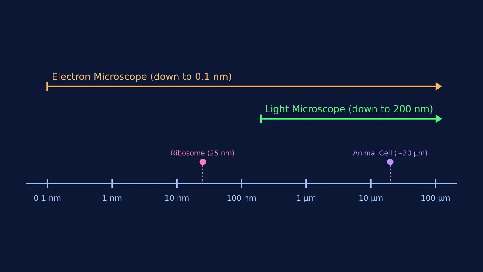
In a standard optical microscope, resolution is bounded by the wavelength of visible light. Because light behaves as a wave, it cannot resolve details much smaller than half its wavelength. This physical boundary restricts light microscopes to a resolution of about 200 nm (Wikipedia). For a deep dive into the mathematics of this limit, you can explore the story of Ernst Abbe and the numerical aperture of the microscope.
Electron microscopes bypass this barrier by using a beam of electrons instead of light. Because the wavelength of an electron is thousands of times shorter than that of visible light, an electron microscope achieves a much higher resolution of about 0.1 nm (Wikipedia).
Why do ribosomes and the ER need an electron microscope?
To understand why cell biologists rely on electron beams, you have to look at the numbers. Eukaryotic ribosomes are between 25 and 30 nm in diameter, while bacterial ribosomes are slightly smaller at 20 nm (Wikipedia).
Because these protein factories are roughly ten times smaller than the 200 nm limit of visible light, a light microscope simply cannot resolve them; the light waves wash right over them. This is also why the detailed folds of the endoplasmic reticulum (ER) remain hidden. The rough ER is densely studded with ribosomes. To see these studs and the delicate membranous tubules, scientists had to wait for the electron microscope. It was this leap in resolution that allowed researchers like George Palade to discover the ribosomes in the cell's protein factory.
Which microscope should you use?
For exam questions and practical decisions, use this framework to choose the correct imaging tool:
Observing living processes: Choose a light microscope. Preparing a sample for an electron microscope requires placing it in a vacuum and often coating it in heavy metals, which kills the cell. If you need to watch a white blood cell move or a cell divide, light is your only option.
Observing natural colour: Choose a light microscope. Electrons do not have "colour" in the human sense; electron micrographs are inherently black and white (though they are often artificially coloured later).
Mapping internal organelle structures: Choose a transmission electron microscope (TEM). If the question asks about the internal structure of mitochondria, the arrangement of the 9+2 axoneme in cilia and flagella, or the shape of a virus, you need the 0.1 nm resolution of an electron beam (Wikipedia).
Viewing 3D surface details: Choose a scanning electron microscope (SEM). It bounces electrons off the surface of a specimen to create a highly detailed, three-dimensional topographical map of objects like pollen grains or insect eyes.
How can you visualise these invisible structures?
Memorising the sizes of organelles can feel abstract when you cannot see them for yourself. When you are struggling to picture how these structures fit together, the Concept Library gives you a clear, interactive map of every concept in biology, helping you place tiny organelles into the bigger picture of the cell.


