
On this page
In NCERT's Cell chapter, ribosomes get one line of history: they were "first observed under the electron microscope as dense particles by George Palade (1953)" (NCERT Class 11 Biology, Chapter 8). Palade did much more than spot them. He worked out where in the cell secretory proteins are made and the route they take out. This post covers both discoveries, the experiment that traced the route, and the numbers behind it.
Who was George Palade?
His Nobel Prize facts page records that he was born on 19 November 1912 in Iasi, Romania, and died on 7 October 2008 in Del Mar, California. In his Nobel autobiography he explains that his father, a professor of philosophy, hoped he would study philosophy, but he "preferred to deal with tangibles and specifics" and entered medical school in Bucharest in 1930.
After the Second World War he came to the United States in 1946. He heard Albert Claude speak about electron microscopy, and Claude invited him to work at the Rockefeller Institute in New York. There Palade and Keith Porter improved the cutting and fixing of tissue until the electron microscope could show the fine structure of cells clearly.
What did he see in 1955?
His paper in volume 1 of The Journal of Cell Biology, A small particulate component of the cytoplasm, described "a particulate component of small dimensions (100 to 150 A) and high density" in mammalian and bird cells. In highly specialised cells the particles were mostly attached to the membrane of the endoplasmic reticulum. In fast-dividing cells they lay more freely in the cytoplasm. The Nobel facts page dates his discovery of ribosomes to 1955, while NCERT gives 1953. If an exam asks, use NCERT's date; the full description you can read today is the 1955 paper.
With the biochemist Philip Siekevitz, Palade then showed that Claude's "microsomes" were fragments of the endoplasmic reticulum and that the particles were made of RNA and protein, his autobiography records.
Worked example: how big is a ribosome?
The 1955 paper gives size in ångström units. One ångström is 10⁻¹⁰ m and one nanometre is 10⁻⁹ m, so 10 Å = 1 nm.
100 Å = 10 nm and 150 Å = 15 nm.
NCERT gives a prokaryotic ribosome as about 15 nm by 20 nm. The numbers are in the same range, which is a quick check that you have converted correctly.
Common mistakes:
Adding Svedberg units. NCERT's 80S ribosome has 60S and 40S subunits, and 60 + 40 is not 80. S values measure how fast particles settle in a centrifuge, which depends on shape as well as mass.
Calling ribosomes membrane-bound. NCERT says they "are not surrounded by any membrane".
Crediting Palade with the Golgi complex. NCERT credits Camillo Golgi (1898) with first observing it; see our post on Golgi and Ramón y Cajal.
How did he trace a protein out of the cell?
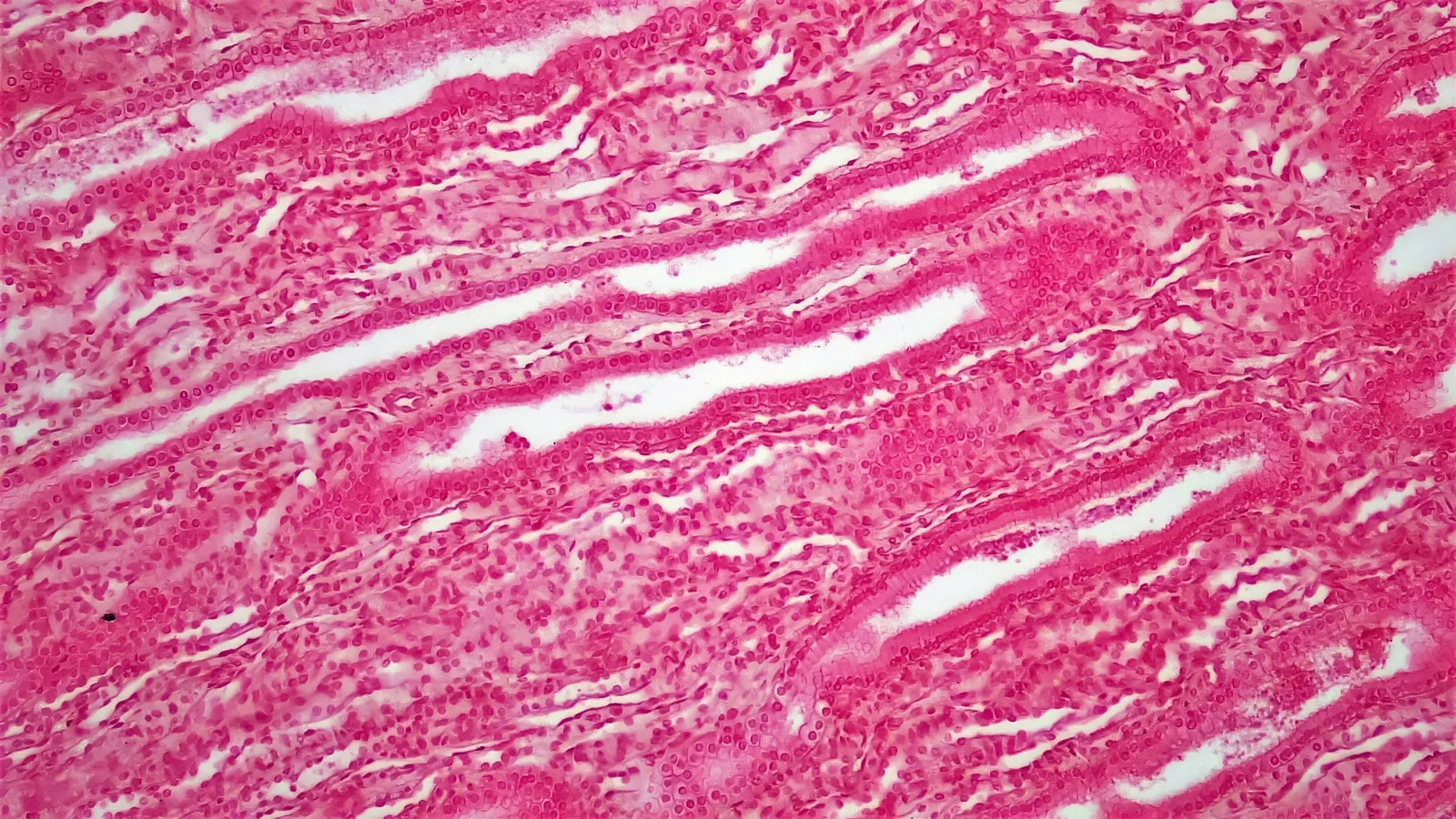
Palade chose the guinea pig pancreas, which makes large amounts of digestive enzymes for export. With James Jamieson he used a pulse-chase method, described in two 1967 papers. First, slices of pancreas were given radioactive leucine for a short pulse, so only proteins made in those minutes carried the label. Then the slices were moved to a "chase" medium with ordinary leucine, and samples were taken at set times to see where the labelled protein had gone.
In the first paper they pulse-labelled for 3 minutes, then chased for a further 7, 17 and 57 minutes. Labelled protein appeared first in rough microsomes, the pieces of rough ER, then moved to smooth microsomes from the edge of the Golgi complex, peaking there at +7 minutes. In the second paper, electron microscope radioautography showed zymogen granules, the storage packets released from the cell, labelled at the earliest at +57 minutes.
Why did this win a Nobel Prize?
The 1974 prize went jointly to Albert Claude, Christian de Duve and Palade, "for their discoveries concerning the structural and functional organization of the cell", with Palade holding a one-third share. His Nobel lecture was titled Intracellular Aspects of the Process of Protein Secretion. The facts page sums up his part: he discovered ribosomes, "where the cell's formation of proteins takes place", and "identified the paths proteins take through the cell".
For what happened to the ribosome after Palade, including its atomic structure, read our post on Venkatraman Ramakrishnan.
What can a student take from his way of working?
Pick the right tissue. A gland that exports protein all day makes the export route easy to see.
Label, then follow. A short pulse and timed samples turned a still picture into a sequence.
Use two methods. Cell fractionation and radioautography checked each other.
If the route from ER to Golgi is hard to picture, the Fermi Simulation Lab offers interactive simulations for ideas easier to see than read, and the Fermi Concept Library has every concept in biology with its own lesson.
Next step: draw the secretory pathway from ribosome to cell membrane and label each stop with Jamieson and Palade's times.