On this page
In 1924 a young London doctor earned a Doctor of Science degree for a thesis on how the human eye forms before birth. Ida Mann turned it into The Development of the Human Eye, published in Cambridge in 1928 (career summary), and the book remained in print for 40 years. In her late fifties she began a second career in the Australian outback.
Who was Ida Mann?
Ida Caroline Mann was born in London in 1893 and died in Perth, Western Australia, in 1983 (biography). She trained at the London School of Medicine for Women, then the only medical school open to women.
A 2024 American Academy of Ophthalmology profile says her anatomy mentor suggested she study the eye using his laboratory's unique collection of human embryo sections. In 1945 she became the first woman to hold a professorship at Oxford in any subject (English Heritage).
How does the human eye develop in the embryo?
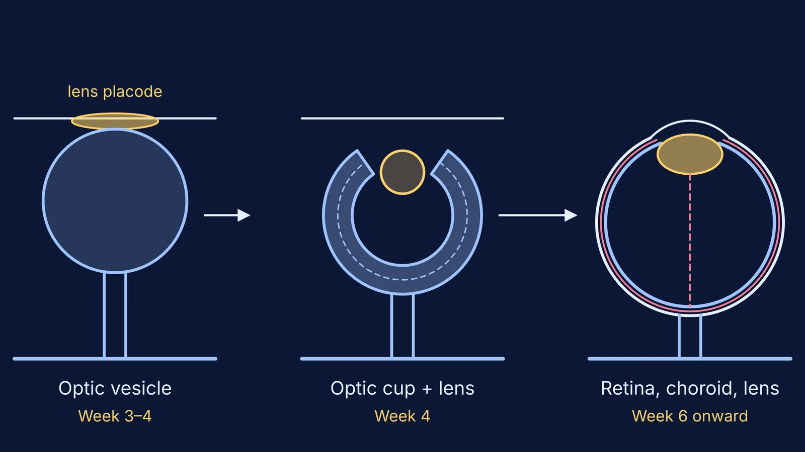
Mann's book follows the eye from its first trace to a recognisable organ. The sequence and timings below are from the NIH-hosted review Embryology, Eye (StatPearls):
Week 3: optic grooves, made of neuroectoderm, appear in the developing forebrain. Shortly after, they bulge out as optic vesicles towards the surface ectoderm.
Next: where a vesicle meets the surface, the ectoderm thickens into a lens placode, which sinks in and pinches off as the lens vesicle.
Week 4: the optic vesicle folds in on itself to form the optic cup, which becomes the retina. A groove beneath it, the choroid fissure, lets the hyaloid artery and vein in.
Weeks 6-7: the choroid, the vascular layer feeding the outer retina, begins to develop. Eyelids form from about week 6, fuse, and separate between the fifth and seventh months.
So the eye has two sources: an outgrowth of the brain gives the retina, and the surface layer gives the lens. If germ layers are hazy, our guide to what organs ectoderm, mesoderm and endoderm form sets out the map.
What did Ida Mann find about the eye's blood vessels?
A 2018 review in Progress in Retinal and Eye Research by Gerard Lutty and D. Scott McLeod cites her 1928 book and says: "The elegant work of Ida Mann suggested that the choroidal vasculature develops first and then the fetal vasculature of vitreous and finally the retinal vasculature."
The same review reports that the hyaloid vessels in the vitreous develop at about 4-6 weeks of gestation and stay intact until about 13 weeks, when they begin to regress. It also disagrees with one of her conclusions, about how those vessels form.
In a 1928 Journal of Anatomy paper she described the hyaloid canal, which runs through the vitreous from the optic nerve to the lens. Our explainer on aqueous and vitreous humour covers what fills the eye after birth.
Her reason, from the preface of her follow-up book, Developmental Abnormalities of the Human Eye, is quoted in the AAO profile: "to evaluate the origin of any developmental anomaly a knowledge of normal growth process is essential."
Why did Ida Mann study trachoma in Western Australia?
Mann moved to Western Australia in 1949 (Encyclopedia of Australian Science and Innovation). After her husband died in 1952, she took on a state eye disease survey around the Kimberley, estimated at about eight weeks; it stretched to four years, by truck on dirt roads and by small plane.
Working in the Kimberley, the north-west coast and the Warburton Ranges, she found trachoma to be the most widespread eye disease among Aboriginal people (Encyclopedia of Women and Leadership in Twentieth-Century Australia). Her findings led to a large treatment programme (EOAS).
Trachoma is an eye disease caused by infection with the bacterium Chlamydia trachomatis, spread by hands, clothes, bedding and flies (WHO). Mann widened the work into a 1966 book, Culture, Race, Climate and Eye Disease, and in 1980 was made a Dame for services to the welfare of Aboriginal people.
WHO later validated India (October 2024) and Australia (April 2026) as having eliminated trachoma as a public health problem.
How can you learn eye development the way Mann saw it?
Mann worked from thin slices of embryos. Try a pencil version:
Draw four frames: optic vesicle, lens placode, optic cup holding the lens vesicle, and an eye with lens and retina.
Colour by origin: one colour for forebrain tissue, another for surface ectoderm.
Write the week under each frame, cover it, and recall it.
Quiz yourself: Which part starts as brain? What passes through the choroid fissure?
Explain the sequence aloud to a friend or parent in one minute.
If one step keeps slipping, the Fermi Concept Library gives each biology concept its own lesson and its place in the map, and Fermi's AI tutor can draw the figure and check your understanding as it goes.
For another scientist who explained the eye, read how Allvar Gullstrand worked out near and far focusing.