
On this page
Your eye is filled with two clear materials, and exam questions love to swap them. The aqueous humour is the thin, watery fluid in front of the lens. The vitreous humour is the clear gel behind it. Both let light through, but they are made, moved and used very differently.
Where are the aqueous and vitreous humour in the eye?
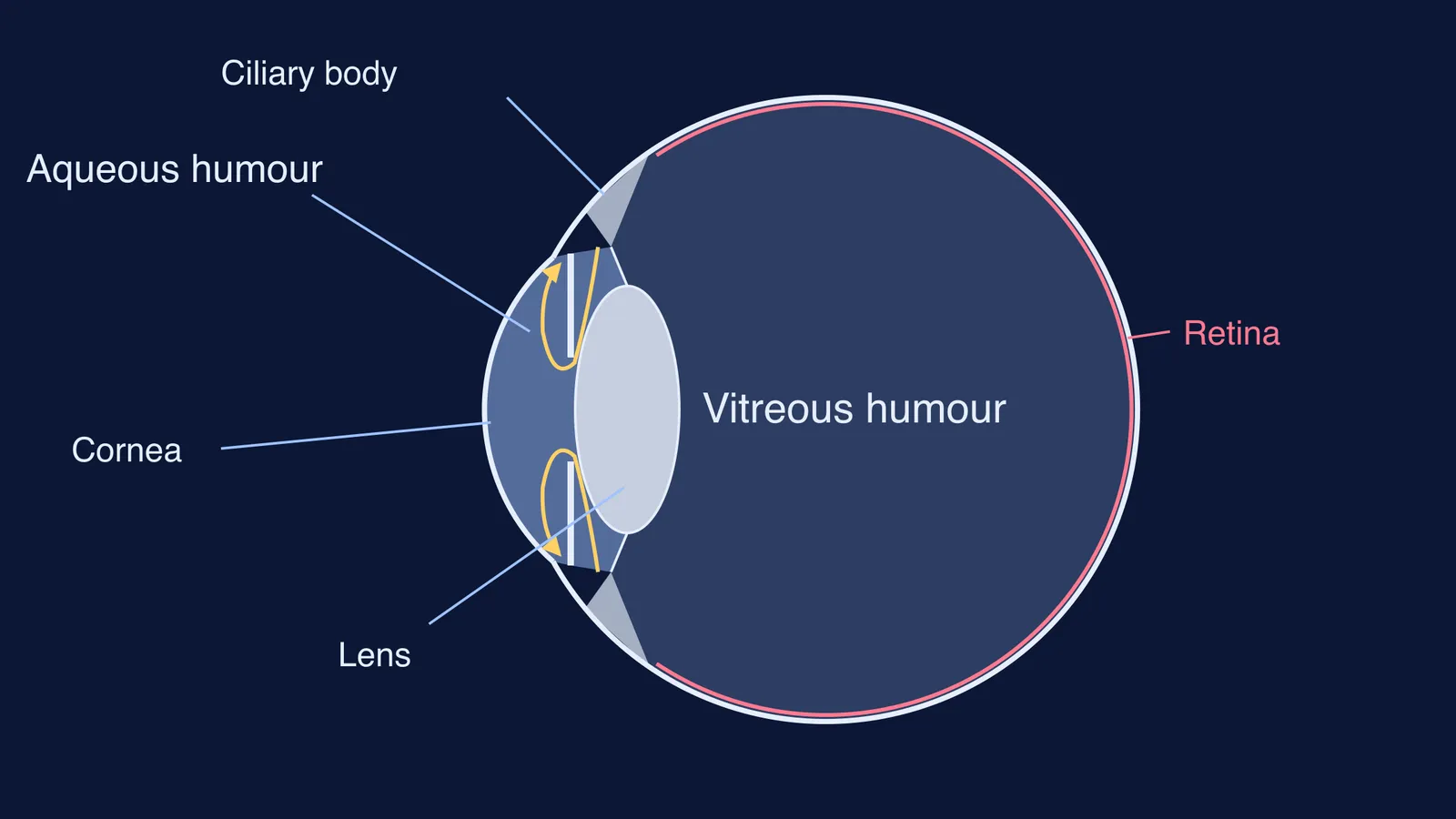
The lens divides the eyeball into a front space and a back space. The front space, between the cornea and the lens, holds the aqueous humour. The iris splits it again: fluid enters the posterior chamber behind the iris, then "flows around the lens and through the pupil into the anterior chamber", as a review of aqueous humour dynamics in The Open Ophthalmology Journal puts it.
The back space, between the lens and the retina, holds the vitreous body. The vitreous occupies about 80% of the globe, so most of the eyeball's volume is gel, not liquid.
What does aqueous humour do?
Aqueous humour is a working fluid that never stops moving. The lens and cornea must stay clear to let light through, so they have no blood vessels; aqueous humour acts as a stand-in for blood, bringing nutrition and carrying away metabolic waste, according to Goel and colleagues' 2010 review.
The ciliary processes, folds of the ciliary body behind the iris, make it. The same review gives a daytime production rate of 2.4 ± 0.6 microlitres per minute in adults.
It leaves the eye by two routes: through the trabecular meshwork into Schlemm's canal, and by a second, uveoscleral route. Because fluid keeps flowing in against resistance on the way out, the eye stays pressurised: an average intraocular pressure of about 15 mmHg in a healthy eye (Goel et al.). The review links raised pressure to glaucoma, a family of diseases that injure the optic nerve.
What does vitreous humour do?
Vitreous humour is a structural gel. It is 99% water, filled with long chains of hyaluronan, with a loose network of type II collagen fibrils in its outer layer, and it has no blood vessels. It helps to keep the retina in position and to maintain the shape of the globe.
It changes slowly over a lifetime. The same Radiopaedia entry notes that liquid pockets inside the gel first appear at around age four, and that shrinking of the vitreous in old age can pull on the retina.
Aqueous vs vitreous humour: the comparison, row by row
Copy these rows into a two-column table in your notes; each compares the same feature on both sides. The facts come from the Open Ophthalmology Journal review and Radiopaedia's vitreous body entry.
Position: aqueous is between the cornea and the lens; vitreous is between the lens and the retina.
Consistency: aqueous is a thin, watery fluid; vitreous is a transparent gel.
Space it fills: aqueous fills the small anterior and posterior chambers; vitreous fills about 80% of the globe.
Made of: aqueous is a clear fluid secreted by the lining of the ciliary processes; vitreous is 99% water with hyaluronan and type II collagen.
Movement: aqueous is made continuously by the ciliary processes and drained through the trabecular meshwork; vitreous is a gel whose liquid part grows slowly with age.
Main job: aqueous feeds the lens and cornea and maintains intraocular pressure; vitreous helps hold the retina in place and keeps the globe's shape.
When it goes wrong: raised intraocular pressure is linked to glaucoma; vitreous shrinkage in old age can pull on the retina.
How do you write the difference in an exam?
For a three-mark "Distinguish between aqueous humour and vitreous humour", give three matched points in this order.
Location: aqueous humour lies in front of the lens, between the cornea and lens; vitreous humour lies behind the lens, between the lens and retina.
Nature: aqueous humour is a thin, watery fluid; vitreous humour is a transparent, jelly-like gel.
Function: aqueous humour nourishes the lens and cornea and maintains intraocular pressure; vitreous humour supports the retina and maintains the shape of the eyeball.
A common slip is writing two unmatched points, such as "aqueous is watery" against "vitreous holds the retina". Pair the features, as in our guide to building a difference table in CBSE biology.
Before you move on, test yourself without looking:
Which humour reaches the anterior chamber through the pupil?
Which one takes up most of the eyeball?
Why can the lens survive without blood vessels?
If any answer felt shaky, the Fermi Concept Library has a lesson for every concept in biology, each with its place in the map, so you can see how the eye's parts connect. Stuck on a diagram question? Fermi's AI tutor can draw the figure and check your understanding as it goes.
The lens that separates the two humours also changes shape to focus; read how in eye accommodation for near and far vision, or see how Kepler showed the retina receives an upside-down image.