On this page
In 1901 a young doctor at Johns Hopkins studied tissue from swollen lymph nodes and asked a simple question: is this disease really tuberculosis in disguise? Dorothy Reed's answer, published in 1902, gave medicine one of its best-known cells, and a way of thinking you can borrow.
Who was Dorothy Reed?
Dorothy Reed Mendenhall (1874-1964) was an American physician who worked in cell pathology, children's medicine and public health, according to the US National Library of Medicine's biography of her. She trained in medicine at Johns Hopkins, and in 1901 she became a pathology fellow there under William Welch (summary of her life on Wikipedia).
What did Dorothy Reed discover about Hodgkin's disease?
The disease was named after Thomas Hodgkin of Guy's Hospital, London, whose 1832 paper described seven patients with painless enlargement of the lymph nodes (Lakhtakia and Burney, Sultan Qaboos University Medical Journal, 2015). Doctors long argued over a basic question: was it an infection, an inflammation or a tumour? Carl Sternberg described and drew its unusual cells in 1898, yet the title of his paper still called the disease a peculiar tuberculosis of the lymphatic system (same source).
Reed tested that idea directly. She compared tissue from tuberculosis patients and from Hodgkin's patients, and in the Hodgkin's tissue saw a large, distinctive cell that the tuberculosis samples lacked, disproving the then-common belief that Hodgkin's was a subtype of tuberculosis (Wikipedia).
Her paper, "On the pathological changes in Hodgkin's disease, with especial reference to its relation to tuberculosis", appeared in volume 10 of the Johns Hopkins Hospital Reports in 1902. The Garrison-Morton medical history bibliography summarises it as describing the growth of endothelial and reticular cells and the formation of "lymphadenoma cells", which it calls "Dorothy Reed's giant cells". These are the cells that now carry her name alongside Sternberg's.
What is a Reed-Sternberg cell?
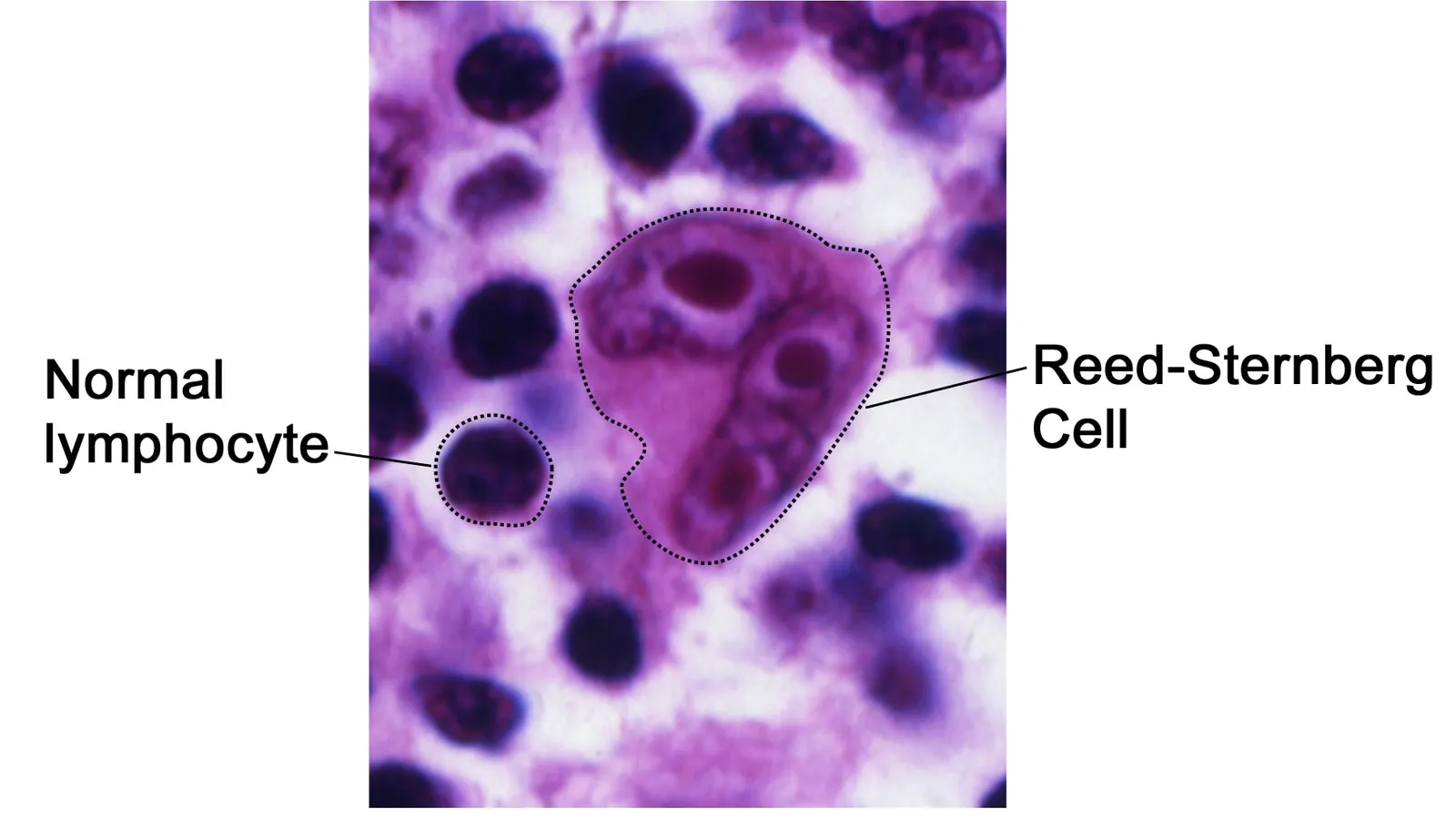
Reed-Sternberg cells are large, abnormal lymphocytes (a type of white blood cell) that may contain more than one nucleus, according to the US National Cancer Institute. Pathologists have nicknamed some forms "owl-eye", "popcorn" and "mirror-image" cells because of the shapes of their nuclei (Lakhtakia and Burney).
Reed could see the cell but not explain it. Chromosome and clonality studies in 1967 and 1975 showed the disease was a true cancer, and in 1999 genetic analysis traced the Reed-Sternberg cell to germinal-centre B cells (same source). Today a pathologist examining a lymph node biopsy looks under the microscope for Reed-Sternberg cells, the National Cancer Institute explains. More than a century on, her observation is still part of the diagnosis.
If lymph and lymphocytes blur together for you, our explainer on how lymph differs from plasma and tissue fluid is a quick refresher, and what makes a tumour benign or malignant covers the cancer side.
Why did Dorothy Reed leave pathology?
Sources differ. One history notes that a woman's prospects in academic pathology at Johns Hopkins were poor at the time (Lakhtakia and Burney). Another says she declined an extended fellowship because of her family's money troubles and took a paediatrics internship in New York (Wikipedia). In 1915 she set up the first infant welfare clinic in Madison, Wisconsin (same source). She later worked for the US Children's Bureau, and in 1926 compared infant and maternal deaths in Denmark with American figures (NLM).
How can you think like Dorothy Reed in biology?
Reed's tool was a careful comparison: take two things people assume are the same, look at both the same way, and find the feature only one has. Use the same four steps on any board or NEET-UG question that asks you to tell two things apart.
Name the claim. "Hodgkin's disease is a kind of tuberculosis."
Set up a fair comparison. Tissue from both diseases, prepared and viewed the same way.
Find the marker. A feature present in one group and absent in the other: here, the large distinctive cell.
State the conclusion and its limit. "These are different diseases" is supported. "This is what causes it" is not; that took most of another century.
Worked example: try it on plant and animal cells. Claim: a cell is a cell. Comparison: onion peel and cheek cells, stained and viewed at the same magnification. Marker: a cell wall and a large central vacuole in the onion cells only. Conclusion: these are different cell types. Limit: one slide tells you what differs, not why.
When you draw what you see, follow the rules for biology microscope drawings. Reed's own drawings of the cells are still reproduced in histories of the disease (Lakhtakia and Burney).
If a comparison falls apart because one of the ideas underneath it is shaky, the Fermi Concept Library has a lesson for every concept in biology, each with its place in the map, so you can fix the gap before the next question.
Next time your textbook names a cell after a person, ask what that person compared to find it.